Mercury »
PDB 1czs-1g4o »
1dbu »
Mercury in PDB 1dbu: Crystal Structure of Cysteinyl-Trna(Pro) Deacylase Protein From H. Influenzae (HI1434)
Protein crystallography data
The structure of Crystal Structure of Cysteinyl-Trna(Pro) Deacylase Protein From H. Influenzae (HI1434), PDB code: 1dbu
was solved by
H.Zhang,
K.Huang,
Z.Li,
O.Herzberg,
Structure 2 Functionproject (S2F),
with X-Ray Crystallography technique. A brief refinement statistics is given in the table below:
| Resolution Low / High (Å) | 20.00 / 1.80 |
| Space group | C 1 2 1 |
| Cell size a, b, c (Å), α, β, γ (°) | 102.570, 42.490, 32.540, 90.00, 104.17, 90.00 |
| R / Rfree (%) | 19.3 / 26.2 |
Mercury Binding Sites:
The binding sites of Mercury atom in the Crystal Structure of Cysteinyl-Trna(Pro) Deacylase Protein From H. Influenzae (HI1434)
(pdb code 1dbu). This binding sites where shown within
5.0 Angstroms radius around Mercury atom.
In total only one binding site of Mercury was determined in the Crystal Structure of Cysteinyl-Trna(Pro) Deacylase Protein From H. Influenzae (HI1434), PDB code: 1dbu:
In total only one binding site of Mercury was determined in the Crystal Structure of Cysteinyl-Trna(Pro) Deacylase Protein From H. Influenzae (HI1434), PDB code: 1dbu:
Mercury binding site 1 out of 1 in 1dbu
Go back to
Mercury binding site 1 out
of 1 in the Crystal Structure of Cysteinyl-Trna(Pro) Deacylase Protein From H. Influenzae (HI1434)
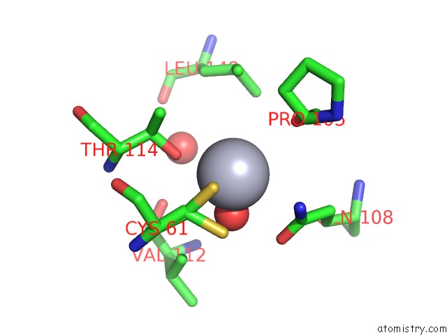
Mono view
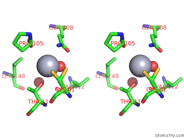
Stereo pair view
Mono view
Stereo pair view
A full contact list of Mercury with other atoms in the Hg binding
site number 1 of Crystal Structure of Cysteinyl-Trna(Pro) Deacylase Protein From H. Influenzae (HI1434) within 5.0Å range:
|
Reference:
H.Zhang,
K.Huang,
Z.Li,
L.Banerjei,
K.E.Fisher,
N.V.Grishin,
E.Eisenstein,
O.Herzberg.
Crystal Structure of Ybak Protein From Haemophilus Influenzae (HI1434) at 1.8 A Resolution: Functional Implications. Proteins V. 40 86 2000.
ISSN: ISSN 0887-3585
PubMed: 10813833
DOI: 10.1002/(SICI)1097-0134(20000701)40:1<86::AID-PROT100>3.0.CO;2-Y
Page generated: Sat Aug 10 23:34:28 2024
ISSN: ISSN 0887-3585
PubMed: 10813833
DOI: 10.1002/(SICI)1097-0134(20000701)40:1<86::AID-PROT100>3.0.CO;2-Y
Last articles
Cl in 7Z0VCl in 7YYG
Cl in 7YWK
Cl in 7YXV
Cl in 7YXM
Cl in 7YUZ
Cl in 7YVV
Cl in 7YWJ
Cl in 7YWB
Cl in 7YRK