Mercury »
PDB 1czs-1g4o »
1dkm »
Mercury in PDB 1dkm: Crystal Structure of Escherichia Coli Phytase at pH 6.6 with HG2+ Cation Acting As An Intermolecular Bridge
Enzymatic activity of Crystal Structure of Escherichia Coli Phytase at pH 6.6 with HG2+ Cation Acting As An Intermolecular Bridge
All present enzymatic activity of Crystal Structure of Escherichia Coli Phytase at pH 6.6 with HG2+ Cation Acting As An Intermolecular Bridge:
3.1.3.2;
3.1.3.2;
Protein crystallography data
The structure of Crystal Structure of Escherichia Coli Phytase at pH 6.6 with HG2+ Cation Acting As An Intermolecular Bridge, PDB code: 1dkm
was solved by
D.Lim,
S.Golovan,
C.W.Forsberg,
Z.Jia,
with X-Ray Crystallography technique. A brief refinement statistics is given in the table below:
| Resolution Low / High (Å) | 25.00 / 2.25 |
| Space group | P 21 21 21 |
| Cell size a, b, c (Å), α, β, γ (°) | 71.026, 74.543, 89.359, 90.00, 90.00, 90.00 |
| R / Rfree (%) | 22.5 / 26.6 |
Mercury Binding Sites:
The binding sites of Mercury atom in the Crystal Structure of Escherichia Coli Phytase at pH 6.6 with HG2+ Cation Acting As An Intermolecular Bridge
(pdb code 1dkm). This binding sites where shown within
5.0 Angstroms radius around Mercury atom.
In total only one binding site of Mercury was determined in the Crystal Structure of Escherichia Coli Phytase at pH 6.6 with HG2+ Cation Acting As An Intermolecular Bridge, PDB code: 1dkm:
In total only one binding site of Mercury was determined in the Crystal Structure of Escherichia Coli Phytase at pH 6.6 with HG2+ Cation Acting As An Intermolecular Bridge, PDB code: 1dkm:
Mercury binding site 1 out of 1 in 1dkm
Go back to
Mercury binding site 1 out
of 1 in the Crystal Structure of Escherichia Coli Phytase at pH 6.6 with HG2+ Cation Acting As An Intermolecular Bridge
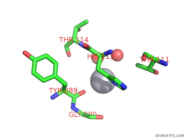
Mono view
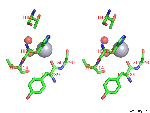
Stereo pair view
Mono view
Stereo pair view
A full contact list of Mercury with other atoms in the Hg binding
site number 1 of Crystal Structure of Escherichia Coli Phytase at pH 6.6 with HG2+ Cation Acting As An Intermolecular Bridge within 5.0Å range:
|
Reference:
D.Lim,
S.Golovan,
C.W.Forsberg,
Z.Jia.
Crystal Structures of Escherichia Coli Phytase and Its Complex with Phytate. Nat.Struct.Biol. V. 7 108 2000.
ISSN: ISSN 1072-8368
PubMed: 10655611
DOI: 10.1038/72371
Page generated: Sat Aug 10 23:34:28 2024
ISSN: ISSN 1072-8368
PubMed: 10655611
DOI: 10.1038/72371
Last articles
Zn in 9JYWZn in 9IR4
Zn in 9IR3
Zn in 9GMX
Zn in 9GMW
Zn in 9JEJ
Zn in 9ERF
Zn in 9ERE
Zn in 9EGV
Zn in 9EGW