Mercury »
PDB 1czs-1g4o »
1dlq »
Mercury in PDB 1dlq: Structure of Catechol 1,2-Dioxygenase From Acinetobacter Sp. ADP1 Inhibited By Bound Mercury
Enzymatic activity of Structure of Catechol 1,2-Dioxygenase From Acinetobacter Sp. ADP1 Inhibited By Bound Mercury
All present enzymatic activity of Structure of Catechol 1,2-Dioxygenase From Acinetobacter Sp. ADP1 Inhibited By Bound Mercury:
1.13.11.1;
1.13.11.1;
Protein crystallography data
The structure of Structure of Catechol 1,2-Dioxygenase From Acinetobacter Sp. ADP1 Inhibited By Bound Mercury, PDB code: 1dlq
was solved by
M.W.Vetting,
D.H.Ohlendorf,
with X-Ray Crystallography technique. A brief refinement statistics is given in the table below:
| Resolution Low / High (Å) | 20.00 / 2.30 |
| Space group | P 1 21 1 |
| Cell size a, b, c (Å), α, β, γ (°) | 52.700, 87.600, 84.000, 90.00, 96.50, 90.00 |
| R / Rfree (%) | 18.8 / 22.4 |
Other elements in 1dlq:
The structure of Structure of Catechol 1,2-Dioxygenase From Acinetobacter Sp. ADP1 Inhibited By Bound Mercury also contains other interesting chemical elements:
| Iron | (Fe) | 2 atoms |
Mercury Binding Sites:
The binding sites of Mercury atom in the Structure of Catechol 1,2-Dioxygenase From Acinetobacter Sp. ADP1 Inhibited By Bound Mercury
(pdb code 1dlq). This binding sites where shown within
5.0 Angstroms radius around Mercury atom.
In total 6 binding sites of Mercury where determined in the Structure of Catechol 1,2-Dioxygenase From Acinetobacter Sp. ADP1 Inhibited By Bound Mercury, PDB code: 1dlq:
Jump to Mercury binding site number: 1; 2; 3; 4; 5; 6;
In total 6 binding sites of Mercury where determined in the Structure of Catechol 1,2-Dioxygenase From Acinetobacter Sp. ADP1 Inhibited By Bound Mercury, PDB code: 1dlq:
Jump to Mercury binding site number: 1; 2; 3; 4; 5; 6;
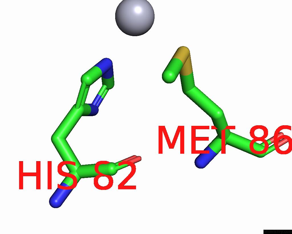
Mercury binding site 1 out of 6 in 1dlq
Go back to
Mercury binding site 1 out
of 6 in the Structure of Catechol 1,2-Dioxygenase From Acinetobacter Sp. ADP1 Inhibited By Bound Mercury

Mono view

Stereo pair view
Mono view
Stereo pair view
A full contact list of Mercury with other atoms in the Hg binding
site number 1 of Structure of Catechol 1,2-Dioxygenase From Acinetobacter Sp. ADP1 Inhibited By Bound Mercury within 5.0Å range:
|
Mercury binding site 2 out of 6 in 1dlq
Go back to
Mercury binding site 2 out
of 6 in the Structure of Catechol 1,2-Dioxygenase From Acinetobacter Sp. ADP1 Inhibited By Bound Mercury

Mono view

Stereo pair view
Mono view
Stereo pair view
A full contact list of Mercury with other atoms in the Hg binding
site number 2 of Structure of Catechol 1,2-Dioxygenase From Acinetobacter Sp. ADP1 Inhibited By Bound Mercury within 5.0Å range:
|
Mercury binding site 3 out of 6 in 1dlq
Go back to
Mercury binding site 3 out
of 6 in the Structure of Catechol 1,2-Dioxygenase From Acinetobacter Sp. ADP1 Inhibited By Bound Mercury
Mono view

Stereo pair view
Mono view
Stereo pair view
A full contact list of Mercury with other atoms in the Hg binding
site number 3 of Structure of Catechol 1,2-Dioxygenase From Acinetobacter Sp. ADP1 Inhibited By Bound Mercury within 5.0Å range:
|
Mercury binding site 4 out of 6 in 1dlq
Go back to
Mercury binding site 4 out
of 6 in the Structure of Catechol 1,2-Dioxygenase From Acinetobacter Sp. ADP1 Inhibited By Bound Mercury

Mono view

Stereo pair view
Mono view
Stereo pair view
A full contact list of Mercury with other atoms in the Hg binding
site number 4 of Structure of Catechol 1,2-Dioxygenase From Acinetobacter Sp. ADP1 Inhibited By Bound Mercury within 5.0Å range:
|
Mercury binding site 5 out of 6 in 1dlq
Go back to
Mercury binding site 5 out
of 6 in the Structure of Catechol 1,2-Dioxygenase From Acinetobacter Sp. ADP1 Inhibited By Bound Mercury

Mono view

Stereo pair view
Mono view
Stereo pair view
A full contact list of Mercury with other atoms in the Hg binding
site number 5 of Structure of Catechol 1,2-Dioxygenase From Acinetobacter Sp. ADP1 Inhibited By Bound Mercury within 5.0Å range:
|
Mercury binding site 6 out of 6 in 1dlq
Go back to
Mercury binding site 6 out
of 6 in the Structure of Catechol 1,2-Dioxygenase From Acinetobacter Sp. ADP1 Inhibited By Bound Mercury

Mono view

Stereo pair view
Mono view
Stereo pair view
A full contact list of Mercury with other atoms in the Hg binding
site number 6 of Structure of Catechol 1,2-Dioxygenase From Acinetobacter Sp. ADP1 Inhibited By Bound Mercury within 5.0Å range:
|
Reference:
M.W.Vetting,
D.H.Ohlendorf.
The 1.8 A Crystal Structure of Catechol 1,2-Dioxygenase Reveals A Novel Hydrophobic Helical Zipper As A Subunit Linker. Structure Fold.Des. V. 8 429 2000.
ISSN: ISSN 0969-2126
PubMed: 10801478
DOI: 10.1016/S0969-2126(00)00122-2
Page generated: Sat Aug 10 23:34:27 2024
ISSN: ISSN 0969-2126
PubMed: 10801478
DOI: 10.1016/S0969-2126(00)00122-2
Last articles
Zn in 9MJ5Zn in 9HNW
Zn in 9G0L
Zn in 9FNE
Zn in 9DZN
Zn in 9E0I
Zn in 9D32
Zn in 9DAK
Zn in 8ZXC
Zn in 8ZUF