Mercury »
PDB 1czs-1g4o »
1dr6 »
Mercury in PDB 1dr6: Crystal Structures of Organomercurial-Activated Chicken Liver Dihydrofolate Reductase Complexes
Enzymatic activity of Crystal Structures of Organomercurial-Activated Chicken Liver Dihydrofolate Reductase Complexes
All present enzymatic activity of Crystal Structures of Organomercurial-Activated Chicken Liver Dihydrofolate Reductase Complexes:
1.5.1.3;
1.5.1.3;
Protein crystallography data
The structure of Crystal Structures of Organomercurial-Activated Chicken Liver Dihydrofolate Reductase Complexes, PDB code: 1dr6
was solved by
M.A.Mctigue,
J.F.Davies /Ii,
B.T.Kaufman,
N.-H.Xuong,
J.Kraut,
with X-Ray Crystallography technique. A brief refinement statistics is given in the table below:
| Resolution Low / High (Å) | N/A / 2.40 |
| Space group | C 1 2 1 |
| Cell size a, b, c (Å), α, β, γ (°) | 88.720, 48.740, 63.870, 90.00, 124.70, 90.00 |
| R / Rfree (%) | n/a / n/a |
Other elements in 1dr6:
The structure of Crystal Structures of Organomercurial-Activated Chicken Liver Dihydrofolate Reductase Complexes also contains other interesting chemical elements:
| Calcium | (Ca) | 1 atom |
Mercury Binding Sites:
The binding sites of Mercury atom in the Crystal Structures of Organomercurial-Activated Chicken Liver Dihydrofolate Reductase Complexes
(pdb code 1dr6). This binding sites where shown within
5.0 Angstroms radius around Mercury atom.
In total 2 binding sites of Mercury where determined in the Crystal Structures of Organomercurial-Activated Chicken Liver Dihydrofolate Reductase Complexes, PDB code: 1dr6:
Jump to Mercury binding site number: 1; 2;
In total 2 binding sites of Mercury where determined in the Crystal Structures of Organomercurial-Activated Chicken Liver Dihydrofolate Reductase Complexes, PDB code: 1dr6:
Jump to Mercury binding site number: 1; 2;
Mercury binding site 1 out of 2 in 1dr6
Go back to
Mercury binding site 1 out
of 2 in the Crystal Structures of Organomercurial-Activated Chicken Liver Dihydrofolate Reductase Complexes
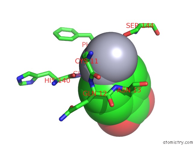
Mono view
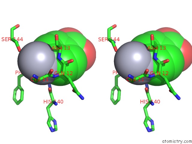
Stereo pair view
Mono view
Stereo pair view
A full contact list of Mercury with other atoms in the Hg binding
site number 1 of Crystal Structures of Organomercurial-Activated Chicken Liver Dihydrofolate Reductase Complexes within 5.0Å range:
|
Mercury binding site 2 out of 2 in 1dr6
Go back to
Mercury binding site 2 out
of 2 in the Crystal Structures of Organomercurial-Activated Chicken Liver Dihydrofolate Reductase Complexes
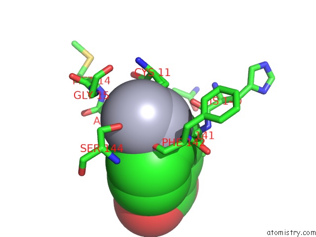
Mono view
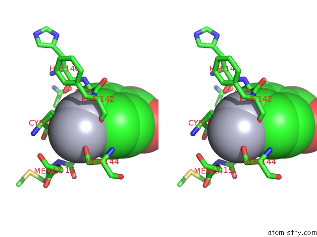
Stereo pair view
Mono view
Stereo pair view
A full contact list of Mercury with other atoms in the Hg binding
site number 2 of Crystal Structures of Organomercurial-Activated Chicken Liver Dihydrofolate Reductase Complexes within 5.0Å range:
|
Reference:
M.A.Mctigue,
J.F.Davies Ii,
B.T.Kaufman,
N.-H.Xuong,
J.Kraut.
Crystal Structures of Organomercurial-Activated Chicken Liver Dihydrofolate Reductase Complexes To Be Published.
Page generated: Fri Aug 8 08:58:12 2025
Last articles
I in 1TUKI in 1TF9
I in 1TB4
I in 1T6H
I in 1T6C
I in 1SD5
I in 1T4E
I in 1SO2
I in 1SRS
I in 1S1M