Mercury »
PDB 1czs-1g4o »
1ems »
Mercury in PDB 1ems: Crystal Structure of the C. Elegans Nitfhit Protein
Protein crystallography data
The structure of Crystal Structure of the C. Elegans Nitfhit Protein, PDB code: 1ems
was solved by
H.C.Pace,
S.C.Hodawadekar,
A.Draganescu,
J.Huang,
P.Bieganowski,
Y.Pekarsky,
C.M.Croce,
C.Brenner,
with X-Ray Crystallography technique. A brief refinement statistics is given in the table below:
| Resolution Low / High (Å) | 30.00 / 2.80 |
| Space group | P 21 21 2 |
| Cell size a, b, c (Å), α, β, γ (°) | 68.750, 100.440, 158.650, 90.00, 90.00, 90.00 |
| R / Rfree (%) | 19 / 23.1 |
Other elements in 1ems:
The structure of Crystal Structure of the C. Elegans Nitfhit Protein also contains other interesting chemical elements:
| Sodium | (Na) | 4 atoms |
Mercury Binding Sites:
The binding sites of Mercury atom in the Crystal Structure of the C. Elegans Nitfhit Protein
(pdb code 1ems). This binding sites where shown within
5.0 Angstroms radius around Mercury atom.
In total 5 binding sites of Mercury where determined in the Crystal Structure of the C. Elegans Nitfhit Protein, PDB code: 1ems:
Jump to Mercury binding site number: 1; 2; 3; 4; 5;
In total 5 binding sites of Mercury where determined in the Crystal Structure of the C. Elegans Nitfhit Protein, PDB code: 1ems:
Jump to Mercury binding site number: 1; 2; 3; 4; 5;
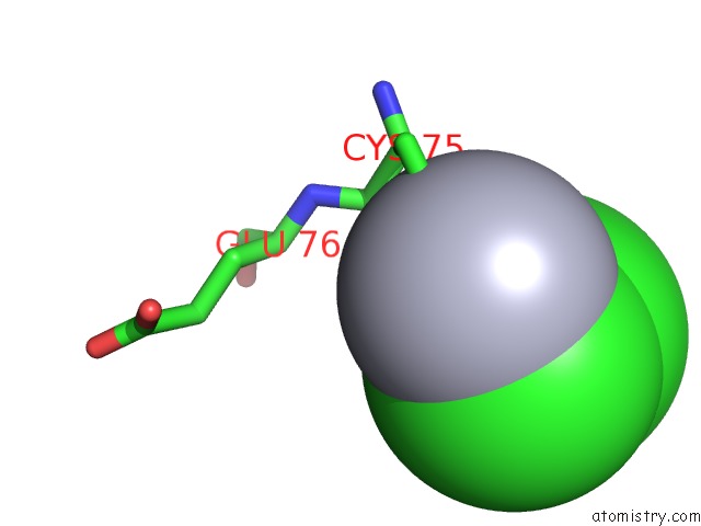
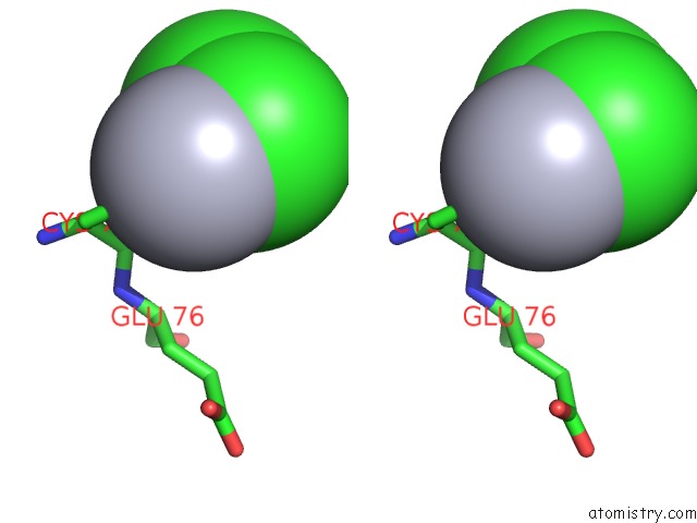
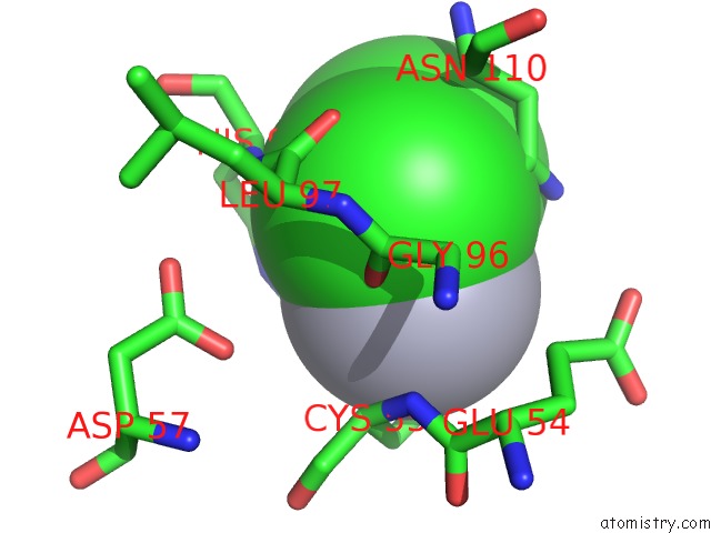
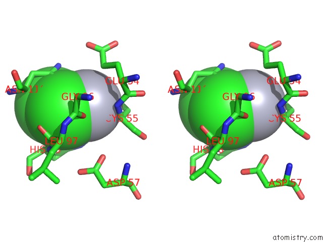
Mercury binding site 1 out of 5 in 1ems
Go back to
Mercury binding site 1 out
of 5 in the Crystal Structure of the C. Elegans Nitfhit Protein
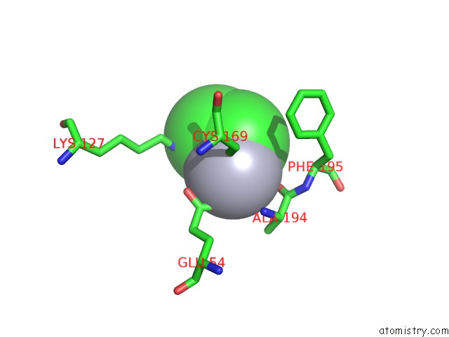
Mono view
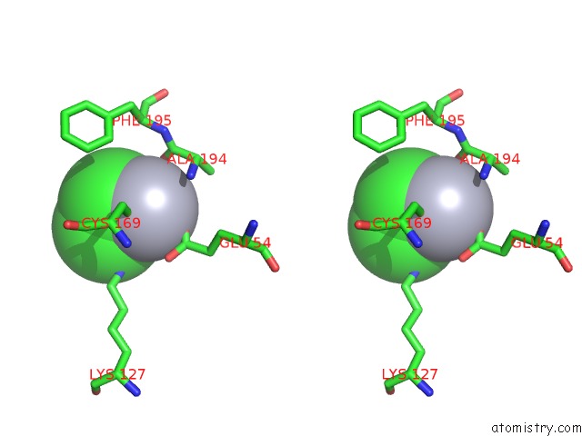
Stereo pair view
Mono view
Stereo pair view
A full contact list of Mercury with other atoms in the Hg binding
site number 1 of Crystal Structure of the C. Elegans Nitfhit Protein within 5.0Å range:
|
Mercury binding site 2 out of 5 in 1ems
Go back to
Mercury binding site 2 out
of 5 in the Crystal Structure of the C. Elegans Nitfhit Protein
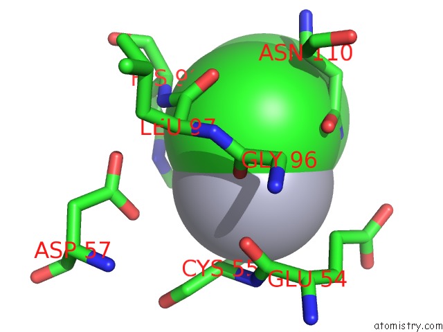
Mono view
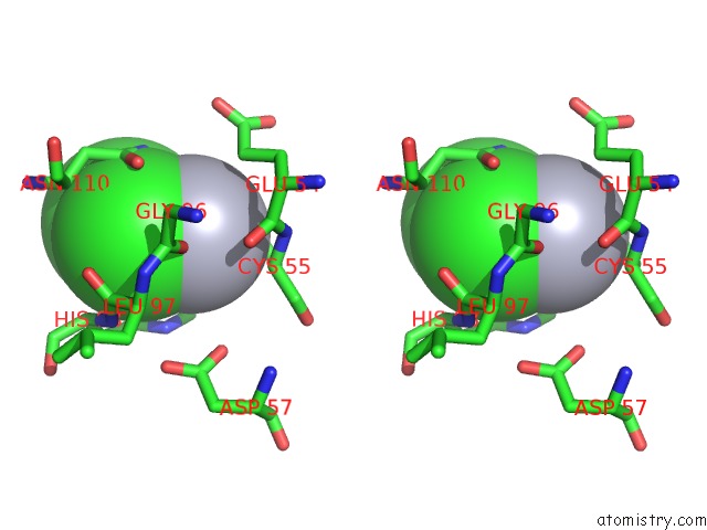
Stereo pair view
Mono view
Stereo pair view
A full contact list of Mercury with other atoms in the Hg binding
site number 2 of Crystal Structure of the C. Elegans Nitfhit Protein within 5.0Å range:
|
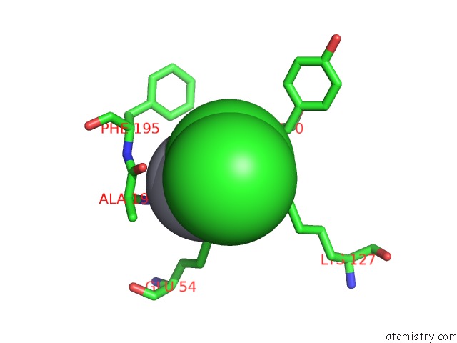
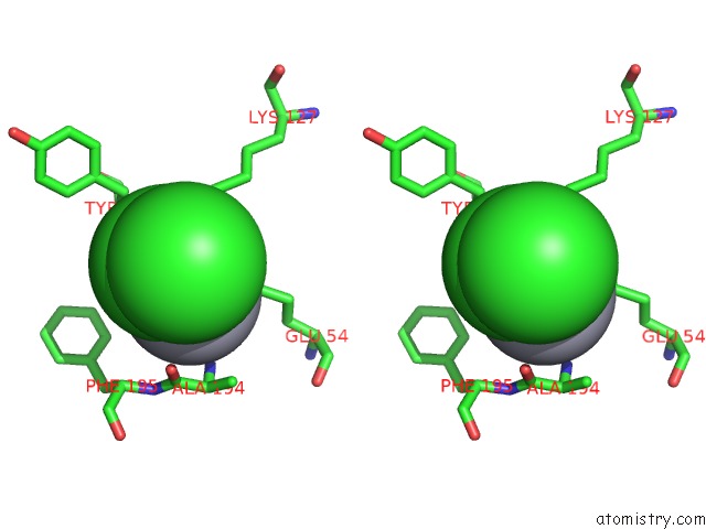
Mercury binding site 3 out of 5 in 1ems
Go back to
Mercury binding site 3 out
of 5 in the Crystal Structure of the C. Elegans Nitfhit Protein
Mono view
Stereo pair view
Mono view
Stereo pair view
A full contact list of Mercury with other atoms in the Hg binding
site number 3 of Crystal Structure of the C. Elegans Nitfhit Protein within 5.0Å range:
|
Mercury binding site 4 out of 5 in 1ems
Go back to
Mercury binding site 4 out
of 5 in the Crystal Structure of the C. Elegans Nitfhit Protein
Mono view
Stereo pair view
Mono view
Stereo pair view
A full contact list of Mercury with other atoms in the Hg binding
site number 4 of Crystal Structure of the C. Elegans Nitfhit Protein within 5.0Å range:
|
Mercury binding site 5 out of 5 in 1ems
Go back to
Mercury binding site 5 out
of 5 in the Crystal Structure of the C. Elegans Nitfhit Protein
Mono view
Stereo pair view
Mono view
Stereo pair view
A full contact list of Mercury with other atoms in the Hg binding
site number 5 of Crystal Structure of the C. Elegans Nitfhit Protein within 5.0Å range:
|
Reference:
H.C.Pace,
S.C.Hodawadekar,
A.Draganescu,
J.Huang,
P.Bieganowski,
Y.Pekarsky,
C.M.Croce,
C.Brenner.
Crystal Structure of the Worm Nitfhit Rosetta Stone Protein Reveals A Nit Tetramer Binding Two Fhit Dimers. Curr.Biol. V. 10 907 2000.
ISSN: ISSN 0960-9822
PubMed: 10959838
DOI: 10.1016/S0960-9822(00)00621-7
Page generated: Sat Aug 10 23:39:55 2024
ISSN: ISSN 0960-9822
PubMed: 10959838
DOI: 10.1016/S0960-9822(00)00621-7
Last articles
F in 7KZ4F in 7KYV
F in 7KYT
F in 7KYB
F in 7KYC
F in 7KYA
F in 7KY5
F in 7KXW
F in 7KY9
F in 7KWA