Mercury »
PDB 1g52-1irk »
1hzx »
Mercury in PDB 1hzx: Crystal Structure of Bovine Rhodopsin
Protein crystallography data
The structure of Crystal Structure of Bovine Rhodopsin, PDB code: 1hzx
was solved by
D.C.Teller,
T.Okada,
C.A.Behnke,
K.Palczewski,
R.E.Stenkamp,
with X-Ray Crystallography technique. A brief refinement statistics is given in the table below:
| Resolution Low / High (Å) | 30.00 / 2.80 |
| Space group | P 41 |
| Cell size a, b, c (Å), α, β, γ (°) | 97.246, 97.246, 149.544, 90.00, 90.00, 90.00 |
| R / Rfree (%) | 17.5 / 21.2 |
Other elements in 1hzx:
The structure of Crystal Structure of Bovine Rhodopsin also contains other interesting chemical elements:
| Zinc | (Zn) | 7 atoms |
Mercury Binding Sites:
The binding sites of Mercury atom in the Crystal Structure of Bovine Rhodopsin
(pdb code 1hzx). This binding sites where shown within
5.0 Angstroms radius around Mercury atom.
In total 6 binding sites of Mercury where determined in the Crystal Structure of Bovine Rhodopsin, PDB code: 1hzx:
Jump to Mercury binding site number: 1; 2; 3; 4; 5; 6;
In total 6 binding sites of Mercury where determined in the Crystal Structure of Bovine Rhodopsin, PDB code: 1hzx:
Jump to Mercury binding site number: 1; 2; 3; 4; 5; 6;
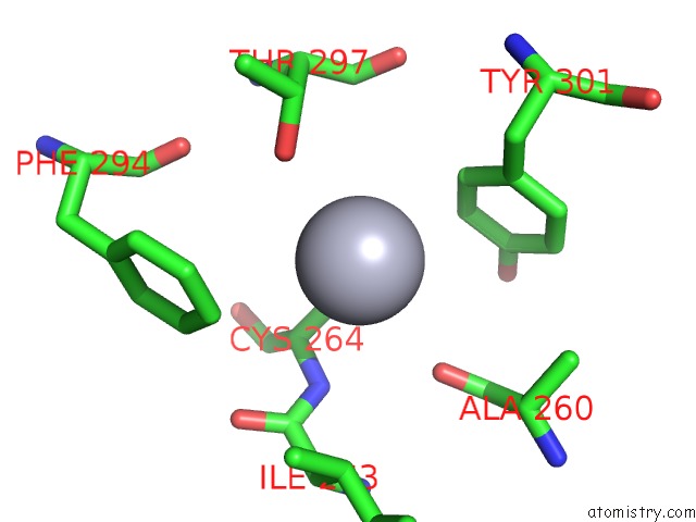
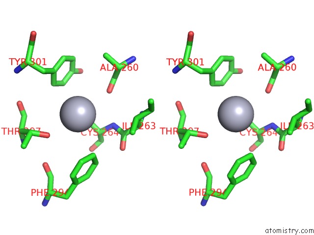
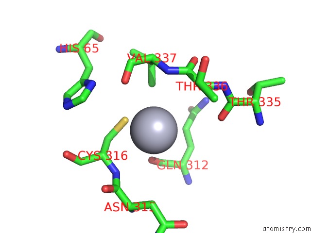
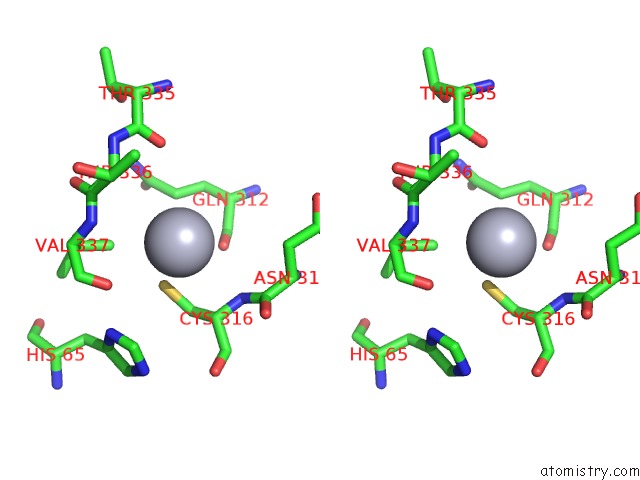
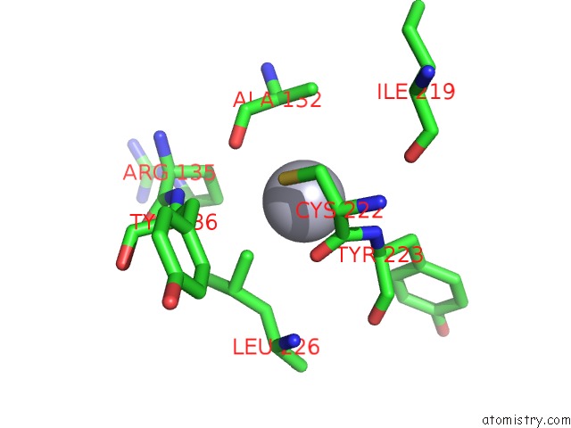
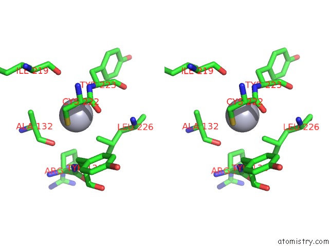
Mercury binding site 1 out of 6 in 1hzx
Go back to
Mercury binding site 1 out
of 6 in the Crystal Structure of Bovine Rhodopsin
Mono view
Stereo pair view
Mono view
Stereo pair view
A full contact list of Mercury with other atoms in the Hg binding
site number 1 of Crystal Structure of Bovine Rhodopsin within 5.0Å range:
|
Mercury binding site 2 out of 6 in 1hzx
Go back to
Mercury binding site 2 out
of 6 in the Crystal Structure of Bovine Rhodopsin
Mono view
Stereo pair view
Mono view
Stereo pair view
A full contact list of Mercury with other atoms in the Hg binding
site number 2 of Crystal Structure of Bovine Rhodopsin within 5.0Å range:
|
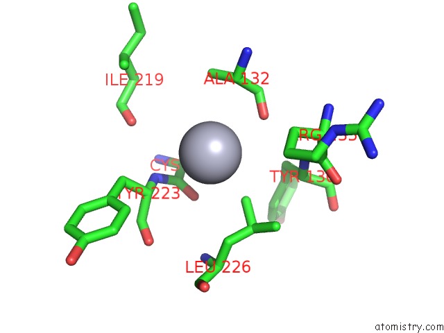
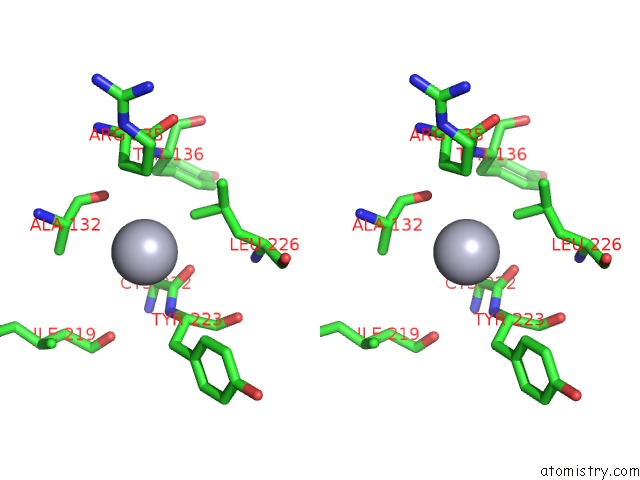
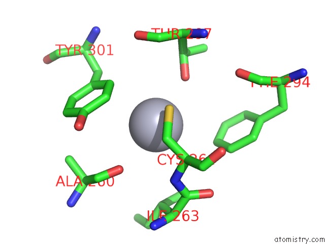
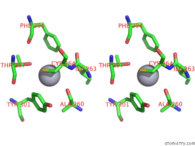
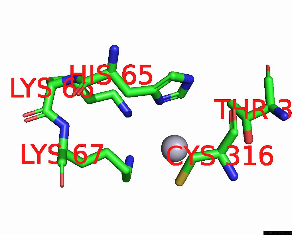
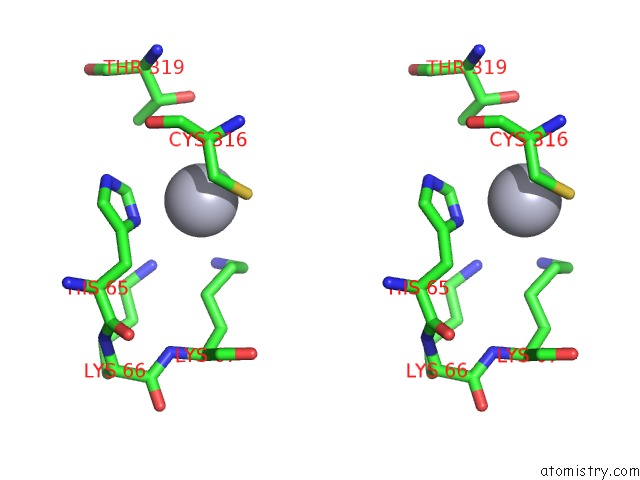
Mercury binding site 3 out of 6 in 1hzx
Go back to
Mercury binding site 3 out
of 6 in the Crystal Structure of Bovine Rhodopsin
Mono view
Stereo pair view
Mono view
Stereo pair view
A full contact list of Mercury with other atoms in the Hg binding
site number 3 of Crystal Structure of Bovine Rhodopsin within 5.0Å range:
|
Mercury binding site 4 out of 6 in 1hzx
Go back to
Mercury binding site 4 out
of 6 in the Crystal Structure of Bovine Rhodopsin
Mono view
Stereo pair view
Mono view
Stereo pair view
A full contact list of Mercury with other atoms in the Hg binding
site number 4 of Crystal Structure of Bovine Rhodopsin within 5.0Å range:
|
Mercury binding site 5 out of 6 in 1hzx
Go back to
Mercury binding site 5 out
of 6 in the Crystal Structure of Bovine Rhodopsin
Mono view
Stereo pair view
Mono view
Stereo pair view
A full contact list of Mercury with other atoms in the Hg binding
site number 5 of Crystal Structure of Bovine Rhodopsin within 5.0Å range:
|
Mercury binding site 6 out of 6 in 1hzx
Go back to
Mercury binding site 6 out
of 6 in the Crystal Structure of Bovine Rhodopsin
Mono view
Stereo pair view
Mono view
Stereo pair view
A full contact list of Mercury with other atoms in the Hg binding
site number 6 of Crystal Structure of Bovine Rhodopsin within 5.0Å range:
|
Reference:
D.C.Teller,
T.Okada,
C.A.Behnke,
K.Palczewski,
R.E.Stenkamp.
Advances in Determination of A High-Resolution Three-Dimensional Structure of Rhodopsin, A Model of G-Protein-Coupled Receptors (Gpcrs). Biochemistry V. 40 7761 2001.
ISSN: ISSN 0006-2960
PubMed: 11425302
DOI: 10.1021/BI0155091
Page generated: Sat Aug 10 23:57:23 2024
ISSN: ISSN 0006-2960
PubMed: 11425302
DOI: 10.1021/BI0155091
Last articles
F in 7NTHF in 7NTI
F in 7NPC
F in 7NRG
F in 7NR5
F in 7NQS
F in 7NOS
F in 7NP5
F in 7NDV
F in 7NP6