Mercury »
PDB 1g52-1irk »
1igw »
Mercury in PDB 1igw: Crystal Structure of the Isocitrate Lyase From the A219C Mutant of Escherichia Coli
Enzymatic activity of Crystal Structure of the Isocitrate Lyase From the A219C Mutant of Escherichia Coli
All present enzymatic activity of Crystal Structure of the Isocitrate Lyase From the A219C Mutant of Escherichia Coli:
4.1.3.1;
4.1.3.1;
Protein crystallography data
The structure of Crystal Structure of the Isocitrate Lyase From the A219C Mutant of Escherichia Coli, PDB code: 1igw
was solved by
K.L.Britton,
I.S.B.Abeysinghe,
P.J.Baker,
V.Barynin,
P.Diehl,
S.J.Langridge,
B.A.Mcfadden,
S.E.Sedelnikova,
T.J.Stillman,
K.Weeradechapon,
D.W.Rice,
with X-Ray Crystallography technique. A brief refinement statistics is given in the table below:
| Resolution Low / High (Å) | 15.00 / 2.10 |
| Space group | P 32 |
| Cell size a, b, c (Å), α, β, γ (°) | 88.650, 88.650, 199.400, 90.00, 90.00, 120.00 |
| R / Rfree (%) | 18.4 / 23.5 |
Other elements in 1igw:
The structure of Crystal Structure of the Isocitrate Lyase From the A219C Mutant of Escherichia Coli also contains other interesting chemical elements:
| Magnesium | (Mg) | 4 atoms |
Mercury Binding Sites:
Pages:
>>> Page 1 <<< Page 2, Binding sites: 11 - 20; Page 3, Binding sites: 21 - 21;Binding sites:
The binding sites of Mercury atom in the Crystal Structure of the Isocitrate Lyase From the A219C Mutant of Escherichia Coli (pdb code 1igw). This binding sites where shown within 5.0 Angstroms radius around Mercury atom.In total 21 binding sites of Mercury where determined in the Crystal Structure of the Isocitrate Lyase From the A219C Mutant of Escherichia Coli, PDB code: 1igw:
Jump to Mercury binding site number: 1; 2; 3; 4; 5; 6; 7; 8; 9; 10;
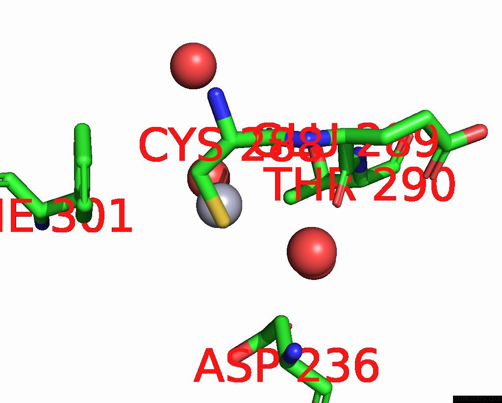
Mercury binding site 1 out of 21 in 1igw
Go back to
Mercury binding site 1 out
of 21 in the Crystal Structure of the Isocitrate Lyase From the A219C Mutant of Escherichia Coli

Mono view

Stereo pair view
Mono view
Stereo pair view
A full contact list of Mercury with other atoms in the Hg binding
site number 1 of Crystal Structure of the Isocitrate Lyase From the A219C Mutant of Escherichia Coli within 5.0Å range:
|
Mercury binding site 2 out of 21 in 1igw
Go back to
Mercury binding site 2 out
of 21 in the Crystal Structure of the Isocitrate Lyase From the A219C Mutant of Escherichia Coli

Mono view

Stereo pair view
Mono view
Stereo pair view
A full contact list of Mercury with other atoms in the Hg binding
site number 2 of Crystal Structure of the Isocitrate Lyase From the A219C Mutant of Escherichia Coli within 5.0Å range:
|
Mercury binding site 3 out of 21 in 1igw
Go back to
Mercury binding site 3 out
of 21 in the Crystal Structure of the Isocitrate Lyase From the A219C Mutant of Escherichia Coli

Mono view

Stereo pair view
Mono view
Stereo pair view
A full contact list of Mercury with other atoms in the Hg binding
site number 3 of Crystal Structure of the Isocitrate Lyase From the A219C Mutant of Escherichia Coli within 5.0Å range:
|
Mercury binding site 4 out of 21 in 1igw
Go back to
Mercury binding site 4 out
of 21 in the Crystal Structure of the Isocitrate Lyase From the A219C Mutant of Escherichia Coli
Mono view

Stereo pair view
Mono view
Stereo pair view
A full contact list of Mercury with other atoms in the Hg binding
site number 4 of Crystal Structure of the Isocitrate Lyase From the A219C Mutant of Escherichia Coli within 5.0Å range:
|
Mercury binding site 5 out of 21 in 1igw
Go back to
Mercury binding site 5 out
of 21 in the Crystal Structure of the Isocitrate Lyase From the A219C Mutant of Escherichia Coli

Mono view

Stereo pair view
Mono view
Stereo pair view
A full contact list of Mercury with other atoms in the Hg binding
site number 5 of Crystal Structure of the Isocitrate Lyase From the A219C Mutant of Escherichia Coli within 5.0Å range:
|
Mercury binding site 6 out of 21 in 1igw
Go back to
Mercury binding site 6 out
of 21 in the Crystal Structure of the Isocitrate Lyase From the A219C Mutant of Escherichia Coli

Mono view

Stereo pair view
Mono view
Stereo pair view
A full contact list of Mercury with other atoms in the Hg binding
site number 6 of Crystal Structure of the Isocitrate Lyase From the A219C Mutant of Escherichia Coli within 5.0Å range:
|
Mercury binding site 7 out of 21 in 1igw
Go back to
Mercury binding site 7 out
of 21 in the Crystal Structure of the Isocitrate Lyase From the A219C Mutant of Escherichia Coli

Mono view

Stereo pair view
Mono view
Stereo pair view
A full contact list of Mercury with other atoms in the Hg binding
site number 7 of Crystal Structure of the Isocitrate Lyase From the A219C Mutant of Escherichia Coli within 5.0Å range:
|
Mercury binding site 8 out of 21 in 1igw
Go back to
Mercury binding site 8 out
of 21 in the Crystal Structure of the Isocitrate Lyase From the A219C Mutant of Escherichia Coli

Mono view

Stereo pair view
Mono view
Stereo pair view
A full contact list of Mercury with other atoms in the Hg binding
site number 8 of Crystal Structure of the Isocitrate Lyase From the A219C Mutant of Escherichia Coli within 5.0Å range:
|
Mercury binding site 9 out of 21 in 1igw
Go back to
Mercury binding site 9 out
of 21 in the Crystal Structure of the Isocitrate Lyase From the A219C Mutant of Escherichia Coli

Mono view

Stereo pair view
Mono view
Stereo pair view
A full contact list of Mercury with other atoms in the Hg binding
site number 9 of Crystal Structure of the Isocitrate Lyase From the A219C Mutant of Escherichia Coli within 5.0Å range:
|
Mercury binding site 10 out of 21 in 1igw
Go back to
Mercury binding site 10 out
of 21 in the Crystal Structure of the Isocitrate Lyase From the A219C Mutant of Escherichia Coli

Mono view

Stereo pair view
Mono view
Stereo pair view
A full contact list of Mercury with other atoms in the Hg binding
site number 10 of Crystal Structure of the Isocitrate Lyase From the A219C Mutant of Escherichia Coli within 5.0Å range:
|
Reference:
K.L.Britton,
I.S.Abeysinghe,
P.J.Baker,
V.Barynin,
P.Diehl,
S.J.Langridge,
B.A.Mcfadden,
S.E.Sedelnikova,
T.J.Stillman,
K.Weeradechapon,
D.W.Rice.
The Structure and Domain Organization of Escherichia Coli Isocitrate Lyase. Acta Crystallogr.,Sect.D V. 57 1209 2001.
ISSN: ISSN 0907-4449
PubMed: 11526312
DOI: 10.1107/S0907444901008642
Page generated: Sun Aug 11 00:04:05 2024
ISSN: ISSN 0907-4449
PubMed: 11526312
DOI: 10.1107/S0907444901008642
Last articles
F in 4CQJF in 4COH
F in 4CNH
F in 4CN4
F in 4CMU
F in 4CMG
F in 4CMB
F in 4CMT
F in 4CM4
F in 4CMO