Mercury »
PDB 1is9-1obh »
1lzj »
Mercury in PDB 1lzj: Glycosyltransferase B + Udp + H Antigen Acceptor
Protein crystallography data
The structure of Glycosyltransferase B + Udp + H Antigen Acceptor, PDB code: 1lzj
was solved by
S.I.Patenaude,
N.O.L.Seto,
S.N.Borisova,
A.Szpacenko,
S.L.Marcus,
M.M.Palcic,
S.V.Evans,
with X-Ray Crystallography technique. A brief refinement statistics is given in the table below:
| Resolution Low / High (Å) | 20.00 / 1.32 |
| Space group | C 2 2 21 |
| Cell size a, b, c (Å), α, β, γ (°) | 52.740, 149.480, 79.530, 90.00, 90.00, 90.00 |
| R / Rfree (%) | 18.4 / 20 |
Other elements in 1lzj:
The structure of Glycosyltransferase B + Udp + H Antigen Acceptor also contains other interesting chemical elements:
| Manganese | (Mn) | 1 atom |
Mercury Binding Sites:
The binding sites of Mercury atom in the Glycosyltransferase B + Udp + H Antigen Acceptor
(pdb code 1lzj). This binding sites where shown within
5.0 Angstroms radius around Mercury atom.
In total 4 binding sites of Mercury where determined in the Glycosyltransferase B + Udp + H Antigen Acceptor, PDB code: 1lzj:
Jump to Mercury binding site number: 1; 2; 3; 4;
In total 4 binding sites of Mercury where determined in the Glycosyltransferase B + Udp + H Antigen Acceptor, PDB code: 1lzj:
Jump to Mercury binding site number: 1; 2; 3; 4;
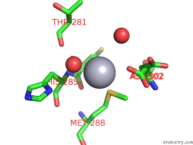
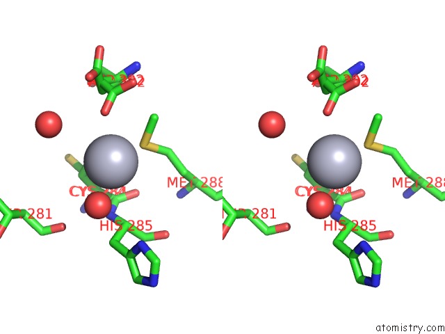
Mercury binding site 1 out of 4 in 1lzj
Go back to
Mercury binding site 1 out
of 4 in the Glycosyltransferase B + Udp + H Antigen Acceptor
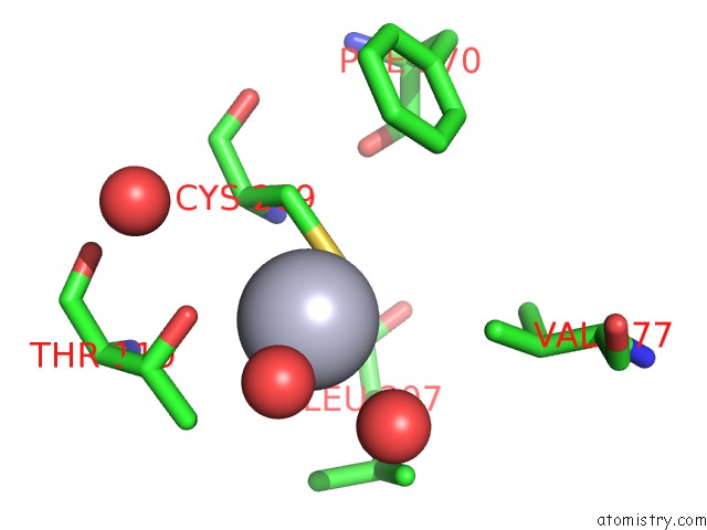
Mono view
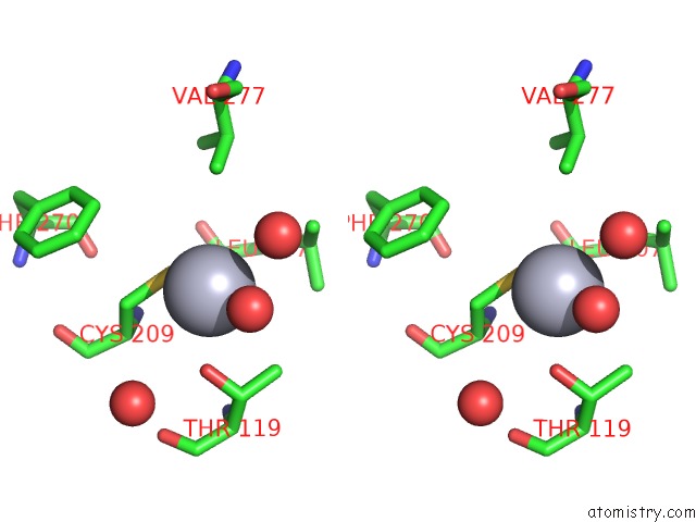
Stereo pair view
Mono view
Stereo pair view
A full contact list of Mercury with other atoms in the Hg binding
site number 1 of Glycosyltransferase B + Udp + H Antigen Acceptor within 5.0Å range:
|
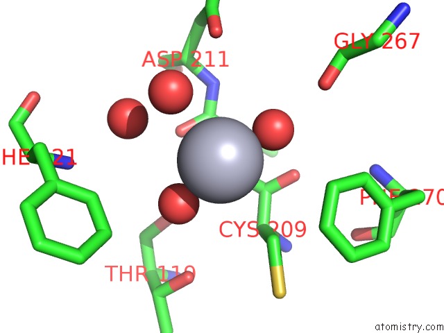
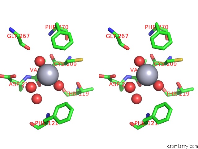
Mercury binding site 2 out of 4 in 1lzj
Go back to
Mercury binding site 2 out
of 4 in the Glycosyltransferase B + Udp + H Antigen Acceptor
Mono view
Stereo pair view
Mono view
Stereo pair view
A full contact list of Mercury with other atoms in the Hg binding
site number 2 of Glycosyltransferase B + Udp + H Antigen Acceptor within 5.0Å range:
|
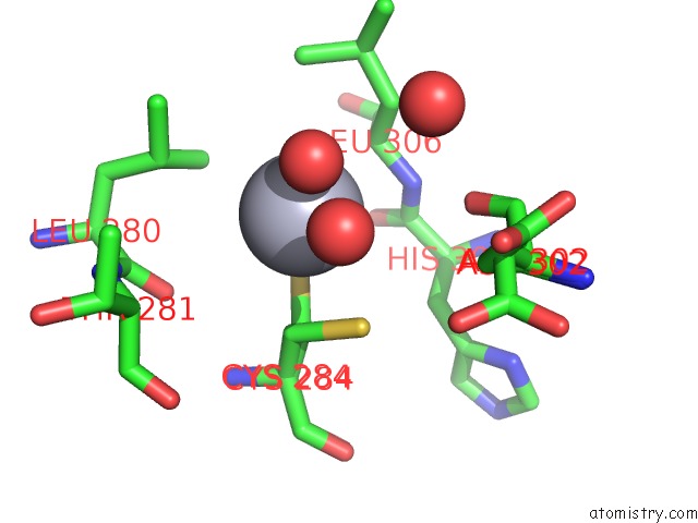
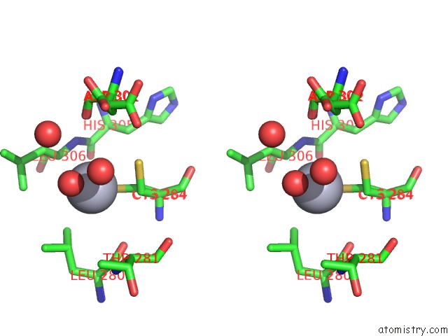
Mercury binding site 3 out of 4 in 1lzj
Go back to
Mercury binding site 3 out
of 4 in the Glycosyltransferase B + Udp + H Antigen Acceptor
Mono view
Stereo pair view
Mono view
Stereo pair view
A full contact list of Mercury with other atoms in the Hg binding
site number 3 of Glycosyltransferase B + Udp + H Antigen Acceptor within 5.0Å range:
|
Mercury binding site 4 out of 4 in 1lzj
Go back to
Mercury binding site 4 out
of 4 in the Glycosyltransferase B + Udp + H Antigen Acceptor
Mono view
Stereo pair view
Mono view
Stereo pair view
A full contact list of Mercury with other atoms in the Hg binding
site number 4 of Glycosyltransferase B + Udp + H Antigen Acceptor within 5.0Å range:
|
Reference:
S.I.Patenaude,
N.O.Seto,
S.N.Borisova,
A.Szpacenko,
S.L.Marcus,
M.M.Palcic,
S.V.Evans.
The Structural Basis For Specificity in Human Abo(H) Blood Group Biosynthesis. Nat.Struct.Biol. V. 9 685 2002.
ISSN: ISSN 1072-8368
PubMed: 12198488
DOI: 10.1038/NSB832
Page generated: Fri Aug 8 09:14:25 2025
ISSN: ISSN 1072-8368
PubMed: 12198488
DOI: 10.1038/NSB832
Last articles
K in 7QQUK in 7QQV
K in 7QQT
K in 7QQR
K in 7QQS
K in 7QQQ
K in 7QQP
K in 7QQO
K in 7QK5
K in 7QIX