Mercury »
PDB 1is9-1obh »
1mrr »
Mercury in PDB 1mrr: Substitution of Manganese For Iron in Ribonucleotide Reductase From Escherichia Coli. Spectroscopic and Crystallographic Characterization
Enzymatic activity of Substitution of Manganese For Iron in Ribonucleotide Reductase From Escherichia Coli. Spectroscopic and Crystallographic Characterization
All present enzymatic activity of Substitution of Manganese For Iron in Ribonucleotide Reductase From Escherichia Coli. Spectroscopic and Crystallographic Characterization:
1.17.4.1;
1.17.4.1;
Protein crystallography data
The structure of Substitution of Manganese For Iron in Ribonucleotide Reductase From Escherichia Coli. Spectroscopic and Crystallographic Characterization, PDB code: 1mrr
was solved by
H.Eklund,
P.Nordlund,
with X-Ray Crystallography technique. A brief refinement statistics is given in the table below:
| Resolution Low / High (Å) | N/A / 2.50 |
| Space group | P 21 21 21 |
| Cell size a, b, c (Å), α, β, γ (°) | 74.300, 85.500, 115.700, 90.00, 90.00, 90.00 |
| R / Rfree (%) | 18 / n/a |
Other elements in 1mrr:
The structure of Substitution of Manganese For Iron in Ribonucleotide Reductase From Escherichia Coli. Spectroscopic and Crystallographic Characterization also contains other interesting chemical elements:
| Manganese | (Mn) | 4 atoms |
Mercury Binding Sites:
Pages:
>>> Page 1 <<< Page 2, Binding sites: 11 - 13;Binding sites:
The binding sites of Mercury atom in the Substitution of Manganese For Iron in Ribonucleotide Reductase From Escherichia Coli. Spectroscopic and Crystallographic Characterization (pdb code 1mrr). This binding sites where shown within 5.0 Angstroms radius around Mercury atom.In total 13 binding sites of Mercury where determined in the Substitution of Manganese For Iron in Ribonucleotide Reductase From Escherichia Coli. Spectroscopic and Crystallographic Characterization, PDB code: 1mrr:
Jump to Mercury binding site number: 1; 2; 3; 4; 5; 6; 7; 8; 9; 10;
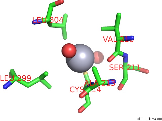
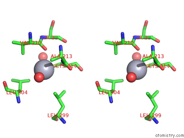
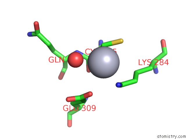
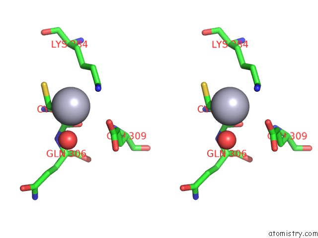
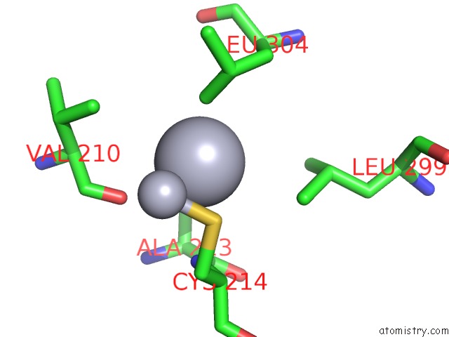
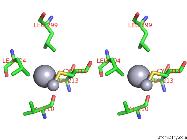
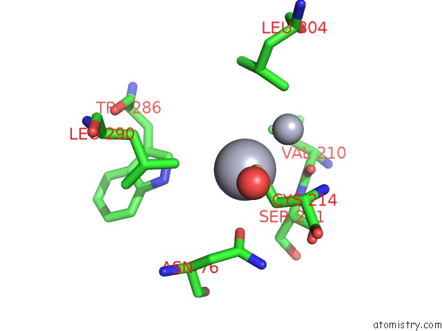
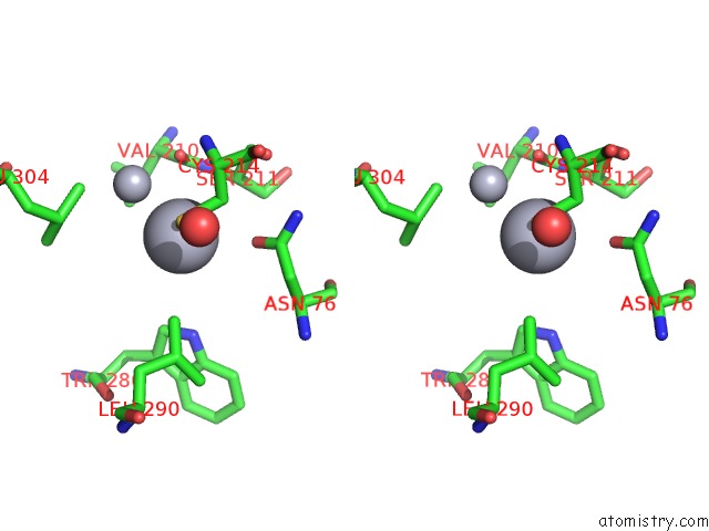
Mercury binding site 1 out of 13 in 1mrr
Go back to
Mercury binding site 1 out
of 13 in the Substitution of Manganese For Iron in Ribonucleotide Reductase From Escherichia Coli. Spectroscopic and Crystallographic Characterization
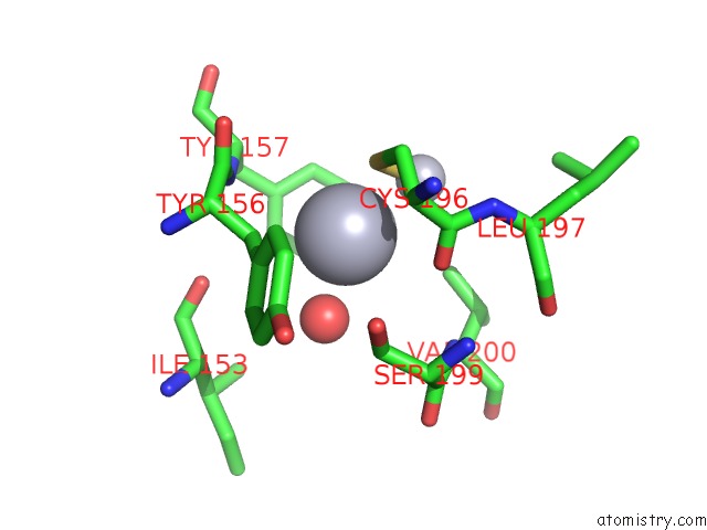
Mono view
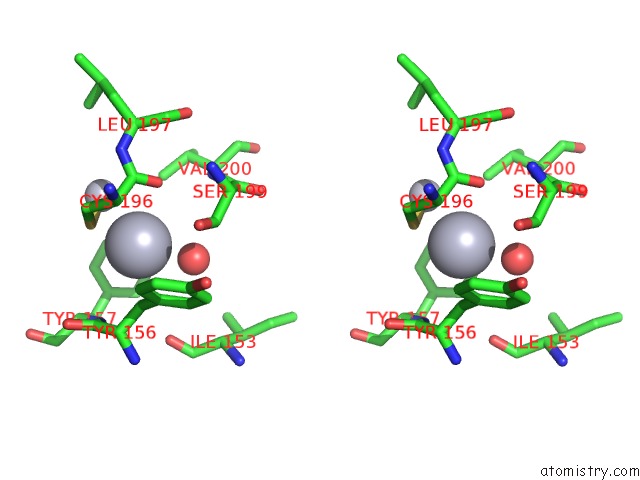
Stereo pair view
Mono view
Stereo pair view
A full contact list of Mercury with other atoms in the Hg binding
site number 1 of Substitution of Manganese For Iron in Ribonucleotide Reductase From Escherichia Coli. Spectroscopic and Crystallographic Characterization within 5.0Å range:
|
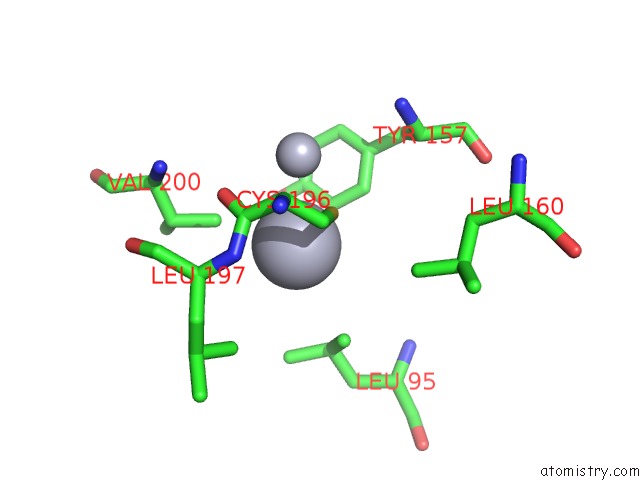
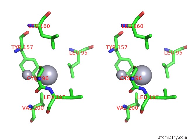
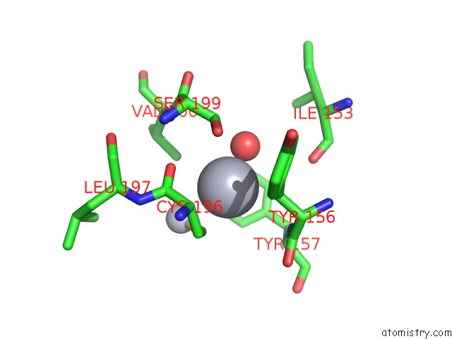
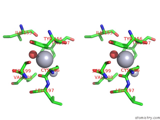
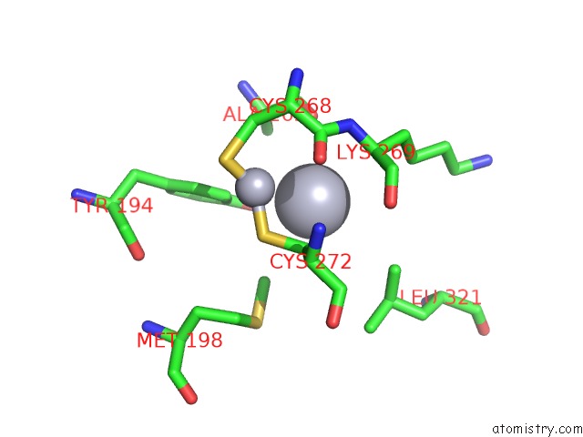
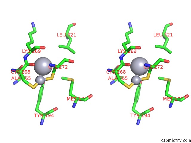
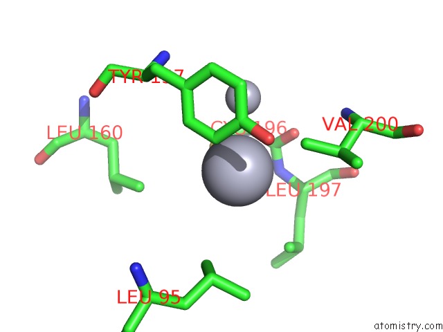
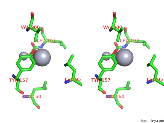
Mercury binding site 2 out of 13 in 1mrr
Go back to
Mercury binding site 2 out
of 13 in the Substitution of Manganese For Iron in Ribonucleotide Reductase From Escherichia Coli. Spectroscopic and Crystallographic Characterization
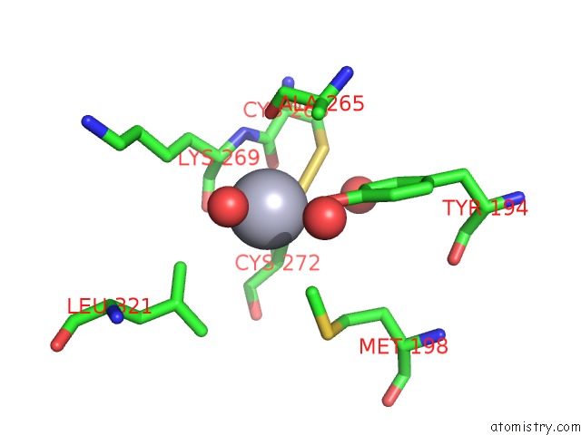
Mono view
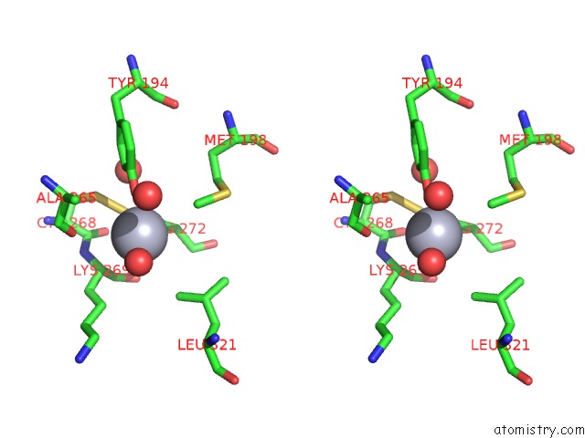
Stereo pair view
Mono view
Stereo pair view
A full contact list of Mercury with other atoms in the Hg binding
site number 2 of Substitution of Manganese For Iron in Ribonucleotide Reductase From Escherichia Coli. Spectroscopic and Crystallographic Characterization within 5.0Å range:
|
Mercury binding site 3 out of 13 in 1mrr
Go back to
Mercury binding site 3 out
of 13 in the Substitution of Manganese For Iron in Ribonucleotide Reductase From Escherichia Coli. Spectroscopic and Crystallographic Characterization
Mono view
Stereo pair view
Mono view
Stereo pair view
A full contact list of Mercury with other atoms in the Hg binding
site number 3 of Substitution of Manganese For Iron in Ribonucleotide Reductase From Escherichia Coli. Spectroscopic and Crystallographic Characterization within 5.0Å range:
|
Mercury binding site 4 out of 13 in 1mrr
Go back to
Mercury binding site 4 out
of 13 in the Substitution of Manganese For Iron in Ribonucleotide Reductase From Escherichia Coli. Spectroscopic and Crystallographic Characterization
Mono view
Stereo pair view
Mono view
Stereo pair view
A full contact list of Mercury with other atoms in the Hg binding
site number 4 of Substitution of Manganese For Iron in Ribonucleotide Reductase From Escherichia Coli. Spectroscopic and Crystallographic Characterization within 5.0Å range:
|
Mercury binding site 5 out of 13 in 1mrr
Go back to
Mercury binding site 5 out
of 13 in the Substitution of Manganese For Iron in Ribonucleotide Reductase From Escherichia Coli. Spectroscopic and Crystallographic Characterization
Mono view
Stereo pair view
Mono view
Stereo pair view
A full contact list of Mercury with other atoms in the Hg binding
site number 5 of Substitution of Manganese For Iron in Ribonucleotide Reductase From Escherichia Coli. Spectroscopic and Crystallographic Characterization within 5.0Å range:
|
Mercury binding site 6 out of 13 in 1mrr
Go back to
Mercury binding site 6 out
of 13 in the Substitution of Manganese For Iron in Ribonucleotide Reductase From Escherichia Coli. Spectroscopic and Crystallographic Characterization
Mono view
Stereo pair view
Mono view
Stereo pair view
A full contact list of Mercury with other atoms in the Hg binding
site number 6 of Substitution of Manganese For Iron in Ribonucleotide Reductase From Escherichia Coli. Spectroscopic and Crystallographic Characterization within 5.0Å range:
|
Mercury binding site 7 out of 13 in 1mrr
Go back to
Mercury binding site 7 out
of 13 in the Substitution of Manganese For Iron in Ribonucleotide Reductase From Escherichia Coli. Spectroscopic and Crystallographic Characterization
Mono view
Stereo pair view
Mono view
Stereo pair view
A full contact list of Mercury with other atoms in the Hg binding
site number 7 of Substitution of Manganese For Iron in Ribonucleotide Reductase From Escherichia Coli. Spectroscopic and Crystallographic Characterization within 5.0Å range:
|
Mercury binding site 8 out of 13 in 1mrr
Go back to
Mercury binding site 8 out
of 13 in the Substitution of Manganese For Iron in Ribonucleotide Reductase From Escherichia Coli. Spectroscopic and Crystallographic Characterization
Mono view
Stereo pair view
Mono view
Stereo pair view
A full contact list of Mercury with other atoms in the Hg binding
site number 8 of Substitution of Manganese For Iron in Ribonucleotide Reductase From Escherichia Coli. Spectroscopic and Crystallographic Characterization within 5.0Å range:
|
Mercury binding site 9 out of 13 in 1mrr
Go back to
Mercury binding site 9 out
of 13 in the Substitution of Manganese For Iron in Ribonucleotide Reductase From Escherichia Coli. Spectroscopic and Crystallographic Characterization
Mono view
Stereo pair view
Mono view
Stereo pair view
A full contact list of Mercury with other atoms in the Hg binding
site number 9 of Substitution of Manganese For Iron in Ribonucleotide Reductase From Escherichia Coli. Spectroscopic and Crystallographic Characterization within 5.0Å range:
|
Mercury binding site 10 out of 13 in 1mrr
Go back to
Mercury binding site 10 out
of 13 in the Substitution of Manganese For Iron in Ribonucleotide Reductase From Escherichia Coli. Spectroscopic and Crystallographic Characterization
Mono view
Stereo pair view
Mono view
Stereo pair view
A full contact list of Mercury with other atoms in the Hg binding
site number 10 of Substitution of Manganese For Iron in Ribonucleotide Reductase From Escherichia Coli. Spectroscopic and Crystallographic Characterization within 5.0Å range:
|
Reference:
M.Atta,
P.Nordlund,
A.Aberg,
H.Eklund,
M.Fontecave.
Substitution of Manganese For Iron in Ribonucleotide Reductase From Escherichia Coli. Spectroscopic and Crystallographic Characterization. J.Biol.Chem. V. 267 20682 1992.
ISSN: ISSN 0021-9258
PubMed: 1328209
Page generated: Fri Aug 8 09:15:40 2025
ISSN: ISSN 0021-9258
PubMed: 1328209
Last articles
I in 4CJDI in 4CDW
I in 4BVX
I in 4BSW
I in 4AW7
I in 4BSV
I in 4B43
I in 4B9H
I in 4AS2
I in 4AS5