Mercury »
PDB 1is9-1obh »
1mxr »
Mercury in PDB 1mxr: High Resolution Structure of Ribonucleotide Reductase R2 From E. Coli in Its Oxidised (Met) Form
Enzymatic activity of High Resolution Structure of Ribonucleotide Reductase R2 From E. Coli in Its Oxidised (Met) Form
All present enzymatic activity of High Resolution Structure of Ribonucleotide Reductase R2 From E. Coli in Its Oxidised (Met) Form:
1.17.4.1;
1.17.4.1;
Protein crystallography data
The structure of High Resolution Structure of Ribonucleotide Reductase R2 From E. Coli in Its Oxidised (Met) Form, PDB code: 1mxr
was solved by
M.A.Andersson,
M.Hogbom,
P.Nordlund,
with X-Ray Crystallography technique. A brief refinement statistics is given in the table below:
| Resolution Low / High (Å) | 36.83 / 1.42 |
| Space group | P 21 21 21 |
| Cell size a, b, c (Å), α, β, γ (°) | 73.844, 84.975, 114.318, 90.00, 90.00, 90.00 |
| R / Rfree (%) | 16 / 18.5 |
Other elements in 1mxr:
The structure of High Resolution Structure of Ribonucleotide Reductase R2 From E. Coli in Its Oxidised (Met) Form also contains other interesting chemical elements:
| Iron | (Fe) | 4 atoms |
Mercury Binding Sites:
Pages:
>>> Page 1 <<< Page 2, Binding sites: 11 - 11;Binding sites:
The binding sites of Mercury atom in the High Resolution Structure of Ribonucleotide Reductase R2 From E. Coli in Its Oxidised (Met) Form (pdb code 1mxr). This binding sites where shown within 5.0 Angstroms radius around Mercury atom.In total 11 binding sites of Mercury where determined in the High Resolution Structure of Ribonucleotide Reductase R2 From E. Coli in Its Oxidised (Met) Form, PDB code: 1mxr:
Jump to Mercury binding site number: 1; 2; 3; 4; 5; 6; 7; 8; 9; 10;
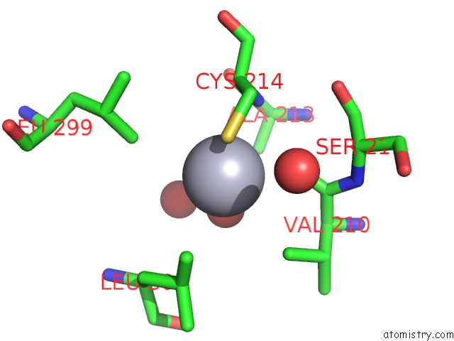
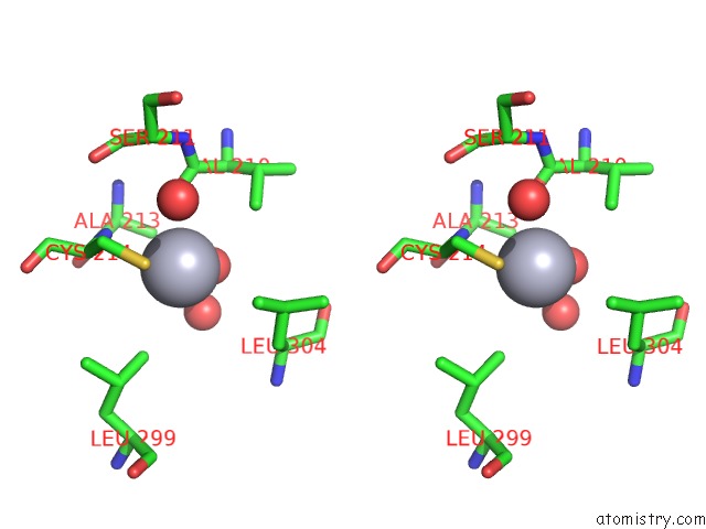
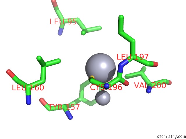
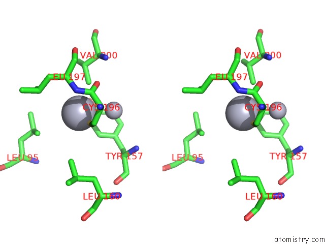
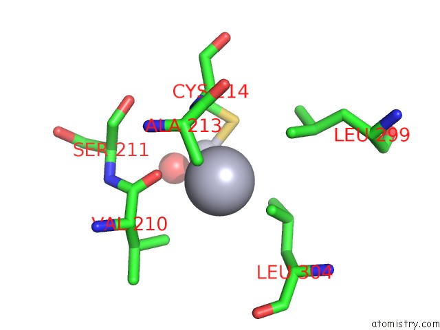
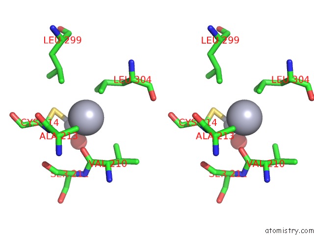
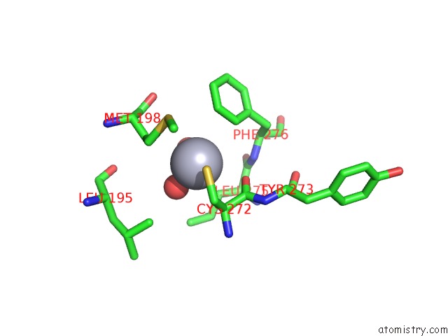
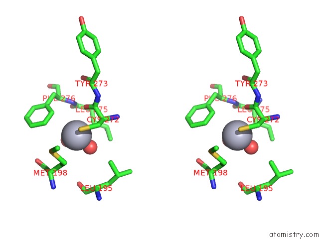
Mercury binding site 1 out of 11 in 1mxr
Go back to
Mercury binding site 1 out
of 11 in the High Resolution Structure of Ribonucleotide Reductase R2 From E. Coli in Its Oxidised (Met) Form
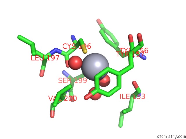
Mono view
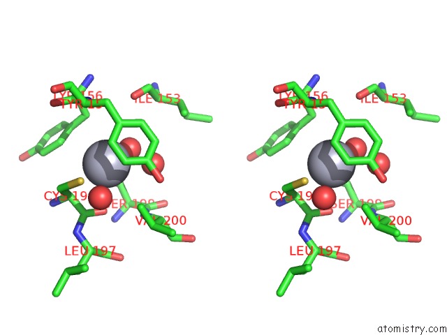
Stereo pair view
Mono view
Stereo pair view
A full contact list of Mercury with other atoms in the Hg binding
site number 1 of High Resolution Structure of Ribonucleotide Reductase R2 From E. Coli in Its Oxidised (Met) Form within 5.0Å range:
|
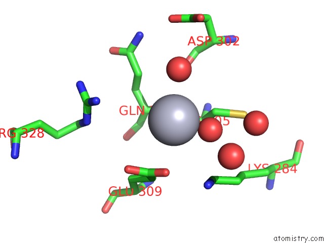
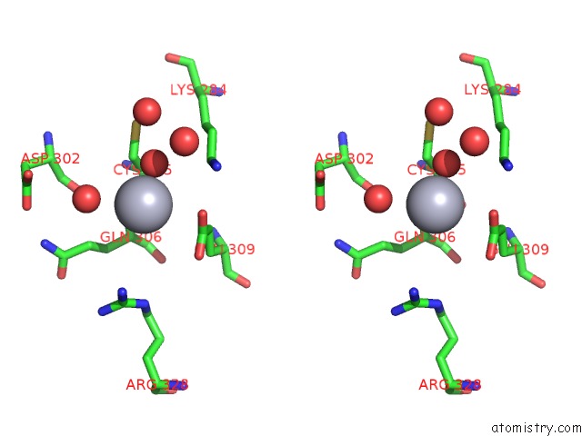
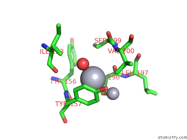
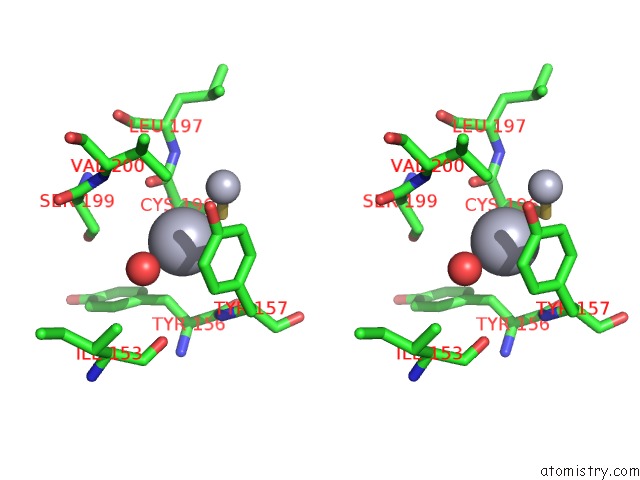
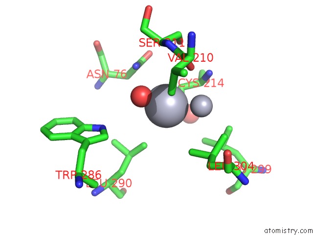
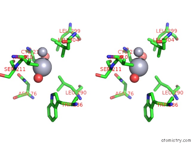
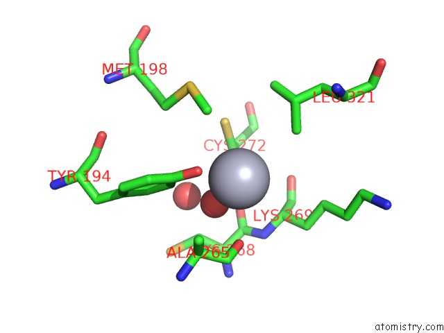
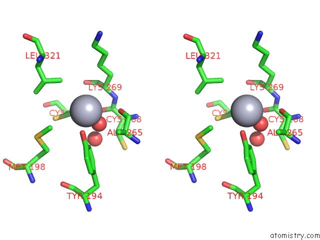
Mercury binding site 2 out of 11 in 1mxr
Go back to
Mercury binding site 2 out
of 11 in the High Resolution Structure of Ribonucleotide Reductase R2 From E. Coli in Its Oxidised (Met) Form
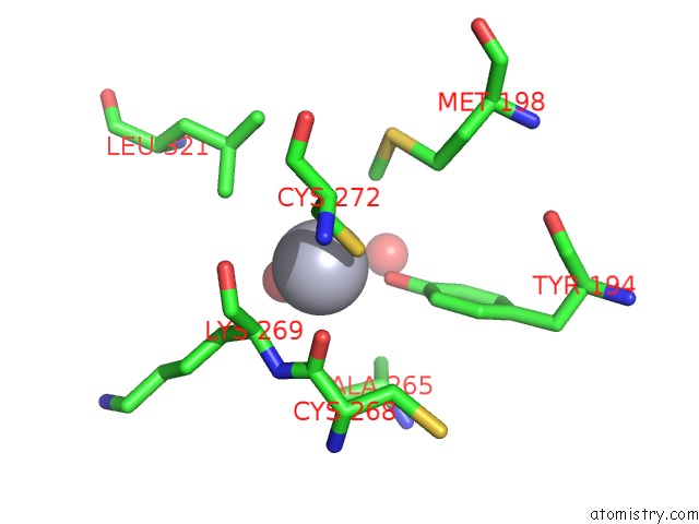
Mono view
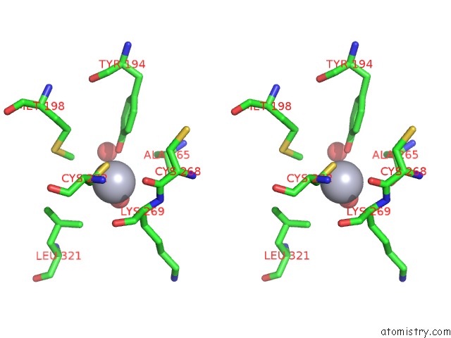
Stereo pair view
Mono view
Stereo pair view
A full contact list of Mercury with other atoms in the Hg binding
site number 2 of High Resolution Structure of Ribonucleotide Reductase R2 From E. Coli in Its Oxidised (Met) Form within 5.0Å range:
|
Mercury binding site 3 out of 11 in 1mxr
Go back to
Mercury binding site 3 out
of 11 in the High Resolution Structure of Ribonucleotide Reductase R2 From E. Coli in Its Oxidised (Met) Form
Mono view
Stereo pair view
Mono view
Stereo pair view
A full contact list of Mercury with other atoms in the Hg binding
site number 3 of High Resolution Structure of Ribonucleotide Reductase R2 From E. Coli in Its Oxidised (Met) Form within 5.0Å range:
|
Mercury binding site 4 out of 11 in 1mxr
Go back to
Mercury binding site 4 out
of 11 in the High Resolution Structure of Ribonucleotide Reductase R2 From E. Coli in Its Oxidised (Met) Form
Mono view
Stereo pair view
Mono view
Stereo pair view
A full contact list of Mercury with other atoms in the Hg binding
site number 4 of High Resolution Structure of Ribonucleotide Reductase R2 From E. Coli in Its Oxidised (Met) Form within 5.0Å range:
|
Mercury binding site 5 out of 11 in 1mxr
Go back to
Mercury binding site 5 out
of 11 in the High Resolution Structure of Ribonucleotide Reductase R2 From E. Coli in Its Oxidised (Met) Form
Mono view
Stereo pair view
Mono view
Stereo pair view
A full contact list of Mercury with other atoms in the Hg binding
site number 5 of High Resolution Structure of Ribonucleotide Reductase R2 From E. Coli in Its Oxidised (Met) Form within 5.0Å range:
|
Mercury binding site 6 out of 11 in 1mxr
Go back to
Mercury binding site 6 out
of 11 in the High Resolution Structure of Ribonucleotide Reductase R2 From E. Coli in Its Oxidised (Met) Form
Mono view
Stereo pair view
Mono view
Stereo pair view
A full contact list of Mercury with other atoms in the Hg binding
site number 6 of High Resolution Structure of Ribonucleotide Reductase R2 From E. Coli in Its Oxidised (Met) Form within 5.0Å range:
|
Mercury binding site 7 out of 11 in 1mxr
Go back to
Mercury binding site 7 out
of 11 in the High Resolution Structure of Ribonucleotide Reductase R2 From E. Coli in Its Oxidised (Met) Form
Mono view
Stereo pair view
Mono view
Stereo pair view
A full contact list of Mercury with other atoms in the Hg binding
site number 7 of High Resolution Structure of Ribonucleotide Reductase R2 From E. Coli in Its Oxidised (Met) Form within 5.0Å range:
|
Mercury binding site 8 out of 11 in 1mxr
Go back to
Mercury binding site 8 out
of 11 in the High Resolution Structure of Ribonucleotide Reductase R2 From E. Coli in Its Oxidised (Met) Form
Mono view
Stereo pair view
Mono view
Stereo pair view
A full contact list of Mercury with other atoms in the Hg binding
site number 8 of High Resolution Structure of Ribonucleotide Reductase R2 From E. Coli in Its Oxidised (Met) Form within 5.0Å range:
|
Mercury binding site 9 out of 11 in 1mxr
Go back to
Mercury binding site 9 out
of 11 in the High Resolution Structure of Ribonucleotide Reductase R2 From E. Coli in Its Oxidised (Met) Form
Mono view
Stereo pair view
Mono view
Stereo pair view
A full contact list of Mercury with other atoms in the Hg binding
site number 9 of High Resolution Structure of Ribonucleotide Reductase R2 From E. Coli in Its Oxidised (Met) Form within 5.0Å range:
|
Mercury binding site 10 out of 11 in 1mxr
Go back to
Mercury binding site 10 out
of 11 in the High Resolution Structure of Ribonucleotide Reductase R2 From E. Coli in Its Oxidised (Met) Form
Mono view
Stereo pair view
Mono view
Stereo pair view
A full contact list of Mercury with other atoms in the Hg binding
site number 10 of High Resolution Structure of Ribonucleotide Reductase R2 From E. Coli in Its Oxidised (Met) Form within 5.0Å range:
|
Reference:
M.Hogbom,
M.Galander,
M.Andersson,
M.Kolberg,
W.Hofbauer,
G.Lassmann,
P.Nordlund,
F.Lendzian.
Displacement of the Tyrosyl Radical Cofactor in Ribonucleotide Reductase Obtained By Single-Crystal High-Field Epr and 1.4-A X-Ray Data. Proc.Natl.Acad.Sci.Usa V. 100 3209 2003.
ISSN: ISSN 0027-8424
PubMed: 12624184
DOI: 10.1073/PNAS.0536684100
Page generated: Sun Aug 11 00:38:49 2024
ISSN: ISSN 0027-8424
PubMed: 12624184
DOI: 10.1073/PNAS.0536684100
Last articles
Zn in 9MJ5Zn in 9HNW
Zn in 9G0L
Zn in 9FNE
Zn in 9DZN
Zn in 9E0I
Zn in 9D32
Zn in 9DAK
Zn in 8ZXC
Zn in 8ZUF