Mercury »
PDB 1of5-1rsr »
1pim »
Mercury in PDB 1pim: Dithionite Reduced E. Coli Ribonucleotide Reductase R2 Subunit, D84E Mutant
Enzymatic activity of Dithionite Reduced E. Coli Ribonucleotide Reductase R2 Subunit, D84E Mutant
All present enzymatic activity of Dithionite Reduced E. Coli Ribonucleotide Reductase R2 Subunit, D84E Mutant:
1.17.4.1;
1.17.4.1;
Protein crystallography data
The structure of Dithionite Reduced E. Coli Ribonucleotide Reductase R2 Subunit, D84E Mutant, PDB code: 1pim
was solved by
W.C.Voegtli,
N.Khidekel,
J.Baldwin,
B.A.Ley,
J.M.Bollinger Jr.,
A.C.Rosenzweig,
with X-Ray Crystallography technique. A brief refinement statistics is given in the table below:
| Resolution Low / High (Å) | 23.75 / 2.00 |
| Space group | P 21 21 21 |
| Cell size a, b, c (Å), α, β, γ (°) | 74.100, 84.600, 114.800, 90.00, 90.00, 90.00 |
| R / Rfree (%) | 20.4 / 24.6 |
Other elements in 1pim:
The structure of Dithionite Reduced E. Coli Ribonucleotide Reductase R2 Subunit, D84E Mutant also contains other interesting chemical elements:
| Iron | (Fe) | 4 atoms |
Mercury Binding Sites:
The binding sites of Mercury atom in the Dithionite Reduced E. Coli Ribonucleotide Reductase R2 Subunit, D84E Mutant
(pdb code 1pim). This binding sites where shown within
5.0 Angstroms radius around Mercury atom.
In total 7 binding sites of Mercury where determined in the Dithionite Reduced E. Coli Ribonucleotide Reductase R2 Subunit, D84E Mutant, PDB code: 1pim:
Jump to Mercury binding site number: 1; 2; 3; 4; 5; 6; 7;
In total 7 binding sites of Mercury where determined in the Dithionite Reduced E. Coli Ribonucleotide Reductase R2 Subunit, D84E Mutant, PDB code: 1pim:
Jump to Mercury binding site number: 1; 2; 3; 4; 5; 6; 7;
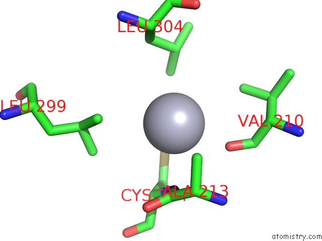
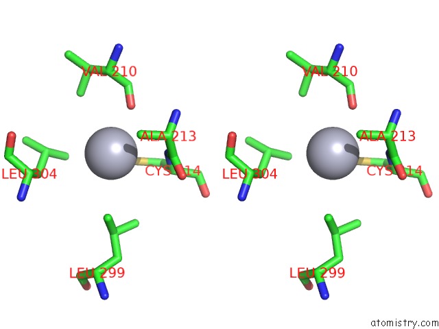
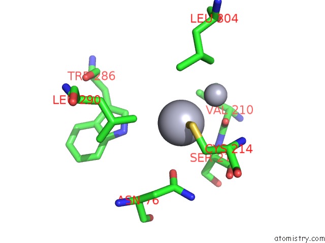
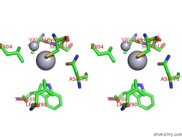
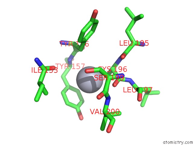
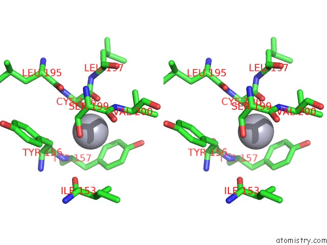
Mercury binding site 1 out of 7 in 1pim
Go back to
Mercury binding site 1 out
of 7 in the Dithionite Reduced E. Coli Ribonucleotide Reductase R2 Subunit, D84E Mutant
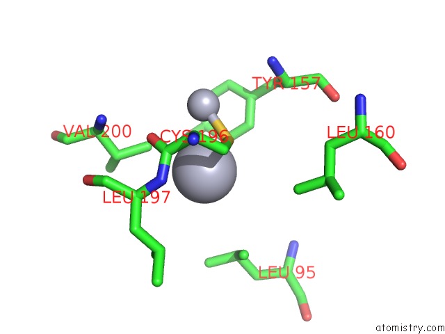
Mono view
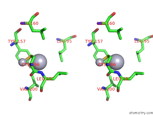
Stereo pair view
Mono view
Stereo pair view
A full contact list of Mercury with other atoms in the Hg binding
site number 1 of Dithionite Reduced E. Coli Ribonucleotide Reductase R2 Subunit, D84E Mutant within 5.0Å range:
|
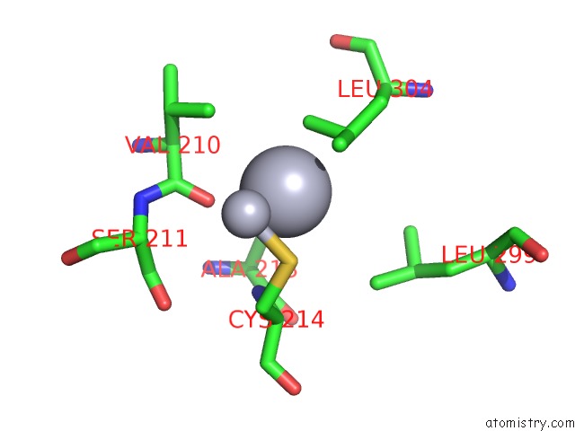
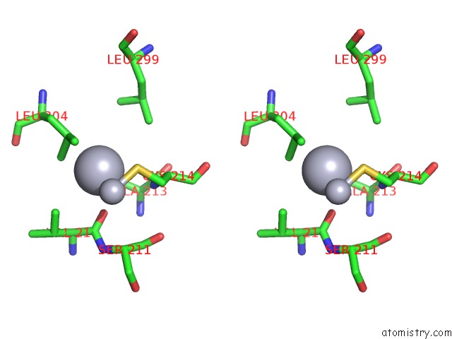
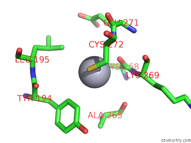
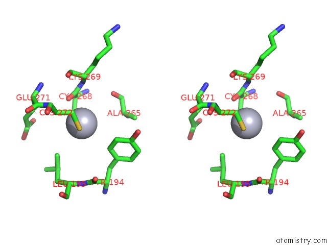
Mercury binding site 2 out of 7 in 1pim
Go back to
Mercury binding site 2 out
of 7 in the Dithionite Reduced E. Coli Ribonucleotide Reductase R2 Subunit, D84E Mutant
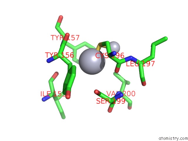
Mono view
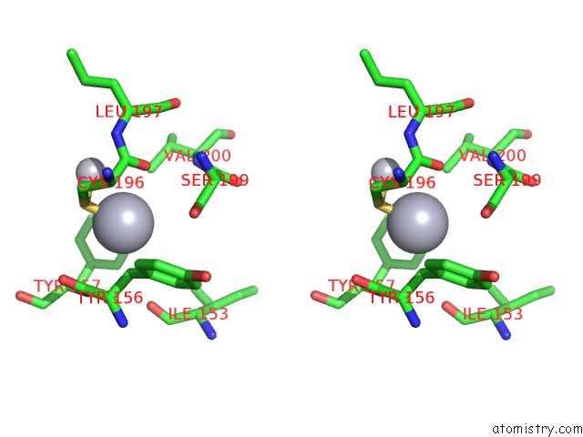
Stereo pair view
Mono view
Stereo pair view
A full contact list of Mercury with other atoms in the Hg binding
site number 2 of Dithionite Reduced E. Coli Ribonucleotide Reductase R2 Subunit, D84E Mutant within 5.0Å range:
|
Mercury binding site 3 out of 7 in 1pim
Go back to
Mercury binding site 3 out
of 7 in the Dithionite Reduced E. Coli Ribonucleotide Reductase R2 Subunit, D84E Mutant
Mono view
Stereo pair view
Mono view
Stereo pair view
A full contact list of Mercury with other atoms in the Hg binding
site number 3 of Dithionite Reduced E. Coli Ribonucleotide Reductase R2 Subunit, D84E Mutant within 5.0Å range:
|
Mercury binding site 4 out of 7 in 1pim
Go back to
Mercury binding site 4 out
of 7 in the Dithionite Reduced E. Coli Ribonucleotide Reductase R2 Subunit, D84E Mutant
Mono view
Stereo pair view
Mono view
Stereo pair view
A full contact list of Mercury with other atoms in the Hg binding
site number 4 of Dithionite Reduced E. Coli Ribonucleotide Reductase R2 Subunit, D84E Mutant within 5.0Å range:
|
Mercury binding site 5 out of 7 in 1pim
Go back to
Mercury binding site 5 out
of 7 in the Dithionite Reduced E. Coli Ribonucleotide Reductase R2 Subunit, D84E Mutant
Mono view
Stereo pair view
Mono view
Stereo pair view
A full contact list of Mercury with other atoms in the Hg binding
site number 5 of Dithionite Reduced E. Coli Ribonucleotide Reductase R2 Subunit, D84E Mutant within 5.0Å range:
|
Mercury binding site 6 out of 7 in 1pim
Go back to
Mercury binding site 6 out
of 7 in the Dithionite Reduced E. Coli Ribonucleotide Reductase R2 Subunit, D84E Mutant
Mono view
Stereo pair view
Mono view
Stereo pair view
A full contact list of Mercury with other atoms in the Hg binding
site number 6 of Dithionite Reduced E. Coli Ribonucleotide Reductase R2 Subunit, D84E Mutant within 5.0Å range:
|
Mercury binding site 7 out of 7 in 1pim
Go back to
Mercury binding site 7 out
of 7 in the Dithionite Reduced E. Coli Ribonucleotide Reductase R2 Subunit, D84E Mutant
Mono view
Stereo pair view
Mono view
Stereo pair view
A full contact list of Mercury with other atoms in the Hg binding
site number 7 of Dithionite Reduced E. Coli Ribonucleotide Reductase R2 Subunit, D84E Mutant within 5.0Å range:
|
Reference:
W.C.Voegtli,
N.Khidekel,
J.Baldwin,
B.A.Ley,
J.M.Bollinger Jr.,
A.C.Rosenzweig.
Crystal Structure of the Ribonucleotide Reductase R2 Mutant That Accumulates A U-1,2-Peroxodiiron(III) Intermediate During Oxygen Activation J.Am.Chem.Soc. V. 122 3255 2000.
ISSN: ISSN 0002-7863
DOI: 10.1021/JA991839L
Page generated: Sun Aug 11 01:00:58 2024
ISSN: ISSN 0002-7863
DOI: 10.1021/JA991839L
Last articles
Zn in 9J0NZn in 9J0O
Zn in 9J0P
Zn in 9FJX
Zn in 9EKB
Zn in 9C0F
Zn in 9CAH
Zn in 9CH0
Zn in 9CH3
Zn in 9CH1