Mercury »
PDB 1of5-1rsr »
1r65 »
Mercury in PDB 1r65: Crystal Structure of Ferrous Soaked Ribonucleotide Reductase R2 Subunit (Wildtype) at pH 5 From E. Coli
Enzymatic activity of Crystal Structure of Ferrous Soaked Ribonucleotide Reductase R2 Subunit (Wildtype) at pH 5 From E. Coli
All present enzymatic activity of Crystal Structure of Ferrous Soaked Ribonucleotide Reductase R2 Subunit (Wildtype) at pH 5 From E. Coli:
1.17.4.1;
1.17.4.1;
Protein crystallography data
The structure of Crystal Structure of Ferrous Soaked Ribonucleotide Reductase R2 Subunit (Wildtype) at pH 5 From E. Coli, PDB code: 1r65
was solved by
W.C.Voegtli,
M.Sommerhalter,
L.Saleh,
J.Baldwin,
J.M.Bollinger Jr.,
A.C.Rosenzweig,
with X-Ray Crystallography technique. A brief refinement statistics is given in the table below:
| Resolution Low / High (Å) | 25.00 / 1.95 |
| Space group | P 21 21 21 |
| Cell size a, b, c (Å), α, β, γ (°) | 74.170, 84.220, 114.220, 90.00, 90.00, 90.00 |
| R / Rfree (%) | 20.7 / 23.6 |
Other elements in 1r65:
The structure of Crystal Structure of Ferrous Soaked Ribonucleotide Reductase R2 Subunit (Wildtype) at pH 5 From E. Coli also contains other interesting chemical elements:
| Iron | (Fe) | 4 atoms |
Mercury Binding Sites:
Pages:
>>> Page 1 <<< Page 2, Binding sites: 11 - 13;Binding sites:
The binding sites of Mercury atom in the Crystal Structure of Ferrous Soaked Ribonucleotide Reductase R2 Subunit (Wildtype) at pH 5 From E. Coli (pdb code 1r65). This binding sites where shown within 5.0 Angstroms radius around Mercury atom.In total 13 binding sites of Mercury where determined in the Crystal Structure of Ferrous Soaked Ribonucleotide Reductase R2 Subunit (Wildtype) at pH 5 From E. Coli, PDB code: 1r65:
Jump to Mercury binding site number: 1; 2; 3; 4; 5; 6; 7; 8; 9; 10;
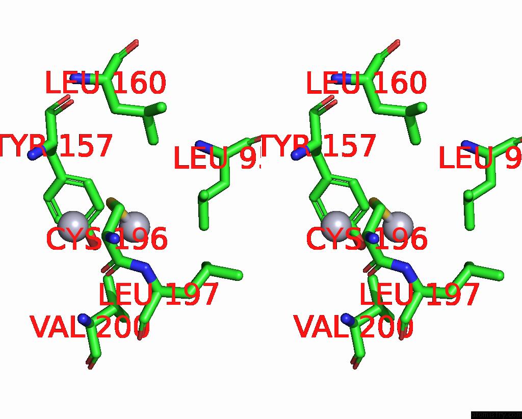
Mercury binding site 1 out of 13 in 1r65
Go back to
Mercury binding site 1 out
of 13 in the Crystal Structure of Ferrous Soaked Ribonucleotide Reductase R2 Subunit (Wildtype) at pH 5 From E. Coli

Mono view

Stereo pair view
Mono view
Stereo pair view
A full contact list of Mercury with other atoms in the Hg binding
site number 1 of Crystal Structure of Ferrous Soaked Ribonucleotide Reductase R2 Subunit (Wildtype) at pH 5 From E. Coli within 5.0Å range:
|
Mercury binding site 2 out of 13 in 1r65
Go back to
Mercury binding site 2 out
of 13 in the Crystal Structure of Ferrous Soaked Ribonucleotide Reductase R2 Subunit (Wildtype) at pH 5 From E. Coli

Mono view

Stereo pair view
Mono view
Stereo pair view
A full contact list of Mercury with other atoms in the Hg binding
site number 2 of Crystal Structure of Ferrous Soaked Ribonucleotide Reductase R2 Subunit (Wildtype) at pH 5 From E. Coli within 5.0Å range:
|
Mercury binding site 3 out of 13 in 1r65
Go back to
Mercury binding site 3 out
of 13 in the Crystal Structure of Ferrous Soaked Ribonucleotide Reductase R2 Subunit (Wildtype) at pH 5 From E. Coli

Mono view

Stereo pair view
Mono view
Stereo pair view
A full contact list of Mercury with other atoms in the Hg binding
site number 3 of Crystal Structure of Ferrous Soaked Ribonucleotide Reductase R2 Subunit (Wildtype) at pH 5 From E. Coli within 5.0Å range:
|
Mercury binding site 4 out of 13 in 1r65
Go back to
Mercury binding site 4 out
of 13 in the Crystal Structure of Ferrous Soaked Ribonucleotide Reductase R2 Subunit (Wildtype) at pH 5 From E. Coli

Mono view

Stereo pair view
Mono view
Stereo pair view
A full contact list of Mercury with other atoms in the Hg binding
site number 4 of Crystal Structure of Ferrous Soaked Ribonucleotide Reductase R2 Subunit (Wildtype) at pH 5 From E. Coli within 5.0Å range:
|
Mercury binding site 5 out of 13 in 1r65
Go back to
Mercury binding site 5 out
of 13 in the Crystal Structure of Ferrous Soaked Ribonucleotide Reductase R2 Subunit (Wildtype) at pH 5 From E. Coli

Mono view
Stereo pair view
Mono view
Stereo pair view
A full contact list of Mercury with other atoms in the Hg binding
site number 5 of Crystal Structure of Ferrous Soaked Ribonucleotide Reductase R2 Subunit (Wildtype) at pH 5 From E. Coli within 5.0Å range:
|
Mercury binding site 6 out of 13 in 1r65
Go back to
Mercury binding site 6 out
of 13 in the Crystal Structure of Ferrous Soaked Ribonucleotide Reductase R2 Subunit (Wildtype) at pH 5 From E. Coli

Mono view

Stereo pair view
Mono view
Stereo pair view
A full contact list of Mercury with other atoms in the Hg binding
site number 6 of Crystal Structure of Ferrous Soaked Ribonucleotide Reductase R2 Subunit (Wildtype) at pH 5 From E. Coli within 5.0Å range:
|
Mercury binding site 7 out of 13 in 1r65
Go back to
Mercury binding site 7 out
of 13 in the Crystal Structure of Ferrous Soaked Ribonucleotide Reductase R2 Subunit (Wildtype) at pH 5 From E. Coli

Mono view

Stereo pair view
Mono view
Stereo pair view
A full contact list of Mercury with other atoms in the Hg binding
site number 7 of Crystal Structure of Ferrous Soaked Ribonucleotide Reductase R2 Subunit (Wildtype) at pH 5 From E. Coli within 5.0Å range:
|
Mercury binding site 8 out of 13 in 1r65
Go back to
Mercury binding site 8 out
of 13 in the Crystal Structure of Ferrous Soaked Ribonucleotide Reductase R2 Subunit (Wildtype) at pH 5 From E. Coli

Mono view

Stereo pair view
Mono view
Stereo pair view
A full contact list of Mercury with other atoms in the Hg binding
site number 8 of Crystal Structure of Ferrous Soaked Ribonucleotide Reductase R2 Subunit (Wildtype) at pH 5 From E. Coli within 5.0Å range:
|
Mercury binding site 9 out of 13 in 1r65
Go back to
Mercury binding site 9 out
of 13 in the Crystal Structure of Ferrous Soaked Ribonucleotide Reductase R2 Subunit (Wildtype) at pH 5 From E. Coli

Mono view

Stereo pair view
Mono view
Stereo pair view
A full contact list of Mercury with other atoms in the Hg binding
site number 9 of Crystal Structure of Ferrous Soaked Ribonucleotide Reductase R2 Subunit (Wildtype) at pH 5 From E. Coli within 5.0Å range:
|
Mercury binding site 10 out of 13 in 1r65
Go back to
Mercury binding site 10 out
of 13 in the Crystal Structure of Ferrous Soaked Ribonucleotide Reductase R2 Subunit (Wildtype) at pH 5 From E. Coli

Mono view

Stereo pair view
Mono view
Stereo pair view
A full contact list of Mercury with other atoms in the Hg binding
site number 10 of Crystal Structure of Ferrous Soaked Ribonucleotide Reductase R2 Subunit (Wildtype) at pH 5 From E. Coli within 5.0Å range:
|
Reference:
W.C.Voegtli,
M.Sommerhalter,
L.Saleh,
J.Baldwin,
J.M.Bollinger Jr.,
A.C.Rosenzweig.
Variable Coordination Geometries at the Diiron(II) Active Site of Ribonucleotide Reductase R2. J.Am.Chem.Soc. V. 125 15822 2003.
ISSN: ISSN 0002-7863
PubMed: 14677973
DOI: 10.1021/JA0370387
Page generated: Fri Aug 8 09:27:50 2025
ISSN: ISSN 0002-7863
PubMed: 14677973
DOI: 10.1021/JA0370387
Last articles
I in 2GSQI in 2GGQ
I in 2FWZ
I in 2G19
I in 2G1M
I in 2DGM
I in 2FWE
I in 2FWF
I in 2FWH
I in 2FDS