Mercury »
PDB 1of5-1rsr »
1r7u »
Mercury in PDB 1r7u: Glycosyltransferase B in Complex with 3-Deoxy-Acceptor Analog Inhibitor
Enzymatic activity of Glycosyltransferase B in Complex with 3-Deoxy-Acceptor Analog Inhibitor
All present enzymatic activity of Glycosyltransferase B in Complex with 3-Deoxy-Acceptor Analog Inhibitor:
2.4.1.37;
2.4.1.37;
Protein crystallography data
The structure of Glycosyltransferase B in Complex with 3-Deoxy-Acceptor Analog Inhibitor, PDB code: 1r7u
was solved by
H.P.Nguyen,
N.O.L.Seto,
Y.Cai,
E.K.Leinala,
S.N.Borisova,
M.M.Palcic,
S.V.Evans,
with X-Ray Crystallography technique. A brief refinement statistics is given in the table below:
| Resolution Low / High (Å) | 19.83 / 1.61 |
| Space group | C 2 2 21 |
| Cell size a, b, c (Å), α, β, γ (°) | 52.600, 149.900, 79.000, 90.00, 90.00, 90.00 |
| R / Rfree (%) | 19.5 / 20.2 |
Mercury Binding Sites:
The binding sites of Mercury atom in the Glycosyltransferase B in Complex with 3-Deoxy-Acceptor Analog Inhibitor
(pdb code 1r7u). This binding sites where shown within
5.0 Angstroms radius around Mercury atom.
In total 4 binding sites of Mercury where determined in the Glycosyltransferase B in Complex with 3-Deoxy-Acceptor Analog Inhibitor, PDB code: 1r7u:
Jump to Mercury binding site number: 1; 2; 3; 4;
In total 4 binding sites of Mercury where determined in the Glycosyltransferase B in Complex with 3-Deoxy-Acceptor Analog Inhibitor, PDB code: 1r7u:
Jump to Mercury binding site number: 1; 2; 3; 4;
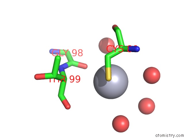
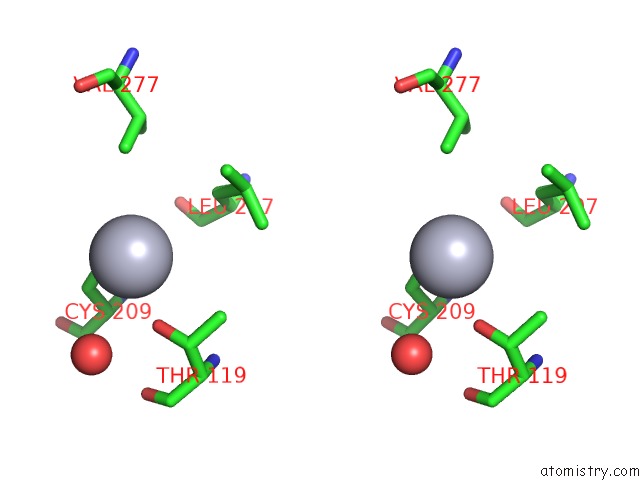
Mercury binding site 1 out of 4 in 1r7u
Go back to
Mercury binding site 1 out
of 4 in the Glycosyltransferase B in Complex with 3-Deoxy-Acceptor Analog Inhibitor
Mono view
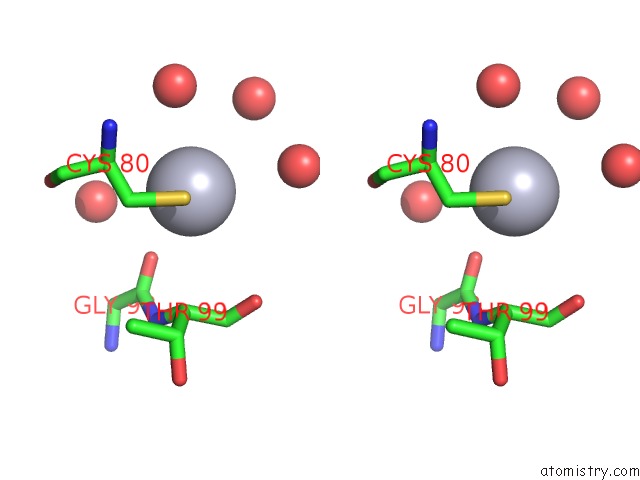
Stereo pair view
Mono view
Stereo pair view
A full contact list of Mercury with other atoms in the Hg binding
site number 1 of Glycosyltransferase B in Complex with 3-Deoxy-Acceptor Analog Inhibitor within 5.0Å range:
|
Mercury binding site 2 out of 4 in 1r7u
Go back to
Mercury binding site 2 out
of 4 in the Glycosyltransferase B in Complex with 3-Deoxy-Acceptor Analog Inhibitor

Mono view
Stereo pair view
Mono view
Stereo pair view
A full contact list of Mercury with other atoms in the Hg binding
site number 2 of Glycosyltransferase B in Complex with 3-Deoxy-Acceptor Analog Inhibitor within 5.0Å range:
|
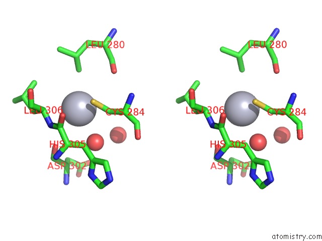
Mercury binding site 3 out of 4 in 1r7u
Go back to
Mercury binding site 3 out
of 4 in the Glycosyltransferase B in Complex with 3-Deoxy-Acceptor Analog Inhibitor
Mono view
Stereo pair view
Mono view
Stereo pair view
A full contact list of Mercury with other atoms in the Hg binding
site number 3 of Glycosyltransferase B in Complex with 3-Deoxy-Acceptor Analog Inhibitor within 5.0Å range:
|
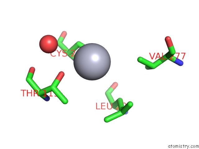
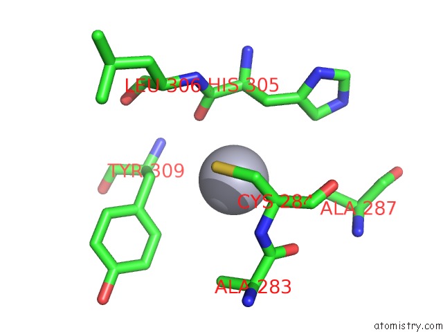
Mercury binding site 4 out of 4 in 1r7u
Go back to
Mercury binding site 4 out
of 4 in the Glycosyltransferase B in Complex with 3-Deoxy-Acceptor Analog Inhibitor
Mono view

Stereo pair view
Mono view
Stereo pair view
A full contact list of Mercury with other atoms in the Hg binding
site number 4 of Glycosyltransferase B in Complex with 3-Deoxy-Acceptor Analog Inhibitor within 5.0Å range:
|
Reference:
H.P.Nguyen,
N.O.L.Seto,
Y.Cai,
E.K.Leinala,
S.N.Borisova,
M.M.Palcic,
S.V.Evans.
The Influence of An Intramolecular Hydrogen Bond in Differential Recognition of Inhibitory Acceptor Analogs By Human Abo(H) Blood Group A and B Glycosyltransferases J.Biol.Chem. V. 278 49191 2003.
ISSN: ISSN 0021-9258
PubMed: 12972418
DOI: 10.1074/JBC.M308770200
Page generated: Sun Aug 11 01:18:22 2024
ISSN: ISSN 0021-9258
PubMed: 12972418
DOI: 10.1074/JBC.M308770200
Last articles
Zn in 9MJ5Zn in 9HNW
Zn in 9G0L
Zn in 9FNE
Zn in 9DZN
Zn in 9E0I
Zn in 9D32
Zn in 9DAK
Zn in 8ZXC
Zn in 8ZUF