Mercury »
PDB 1rsv-1yp2 »
1rwa »
Mercury in PDB 1rwa: Crystal Structure of Arthrobacter Aurescens Chondroitin Ac Lyase
Enzymatic activity of Crystal Structure of Arthrobacter Aurescens Chondroitin Ac Lyase
All present enzymatic activity of Crystal Structure of Arthrobacter Aurescens Chondroitin Ac Lyase:
4.2.2.5;
4.2.2.5;
Protein crystallography data
The structure of Crystal Structure of Arthrobacter Aurescens Chondroitin Ac Lyase, PDB code: 1rwa
was solved by
V.V.Lunin,
Y.Li,
H.Miyazono,
M.Kyogashima,
A.W.Bell,
M.Cygler,
with X-Ray Crystallography technique. A brief refinement statistics is given in the table below:
| Resolution Low / High (Å) | 28.99 / 1.30 |
| Space group | P 1 21 1 |
| Cell size a, b, c (Å), α, β, γ (°) | 57.356, 85.256, 82.162, 90.00, 105.78, 90.00 |
| R / Rfree (%) | 13.4 / 15.6 |
Mercury Binding Sites:
The binding sites of Mercury atom in the Crystal Structure of Arthrobacter Aurescens Chondroitin Ac Lyase
(pdb code 1rwa). This binding sites where shown within
5.0 Angstroms radius around Mercury atom.
In total 3 binding sites of Mercury where determined in the Crystal Structure of Arthrobacter Aurescens Chondroitin Ac Lyase, PDB code: 1rwa:
Jump to Mercury binding site number: 1; 2; 3;
In total 3 binding sites of Mercury where determined in the Crystal Structure of Arthrobacter Aurescens Chondroitin Ac Lyase, PDB code: 1rwa:
Jump to Mercury binding site number: 1; 2; 3;
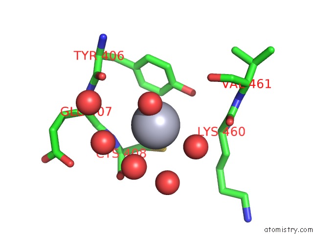
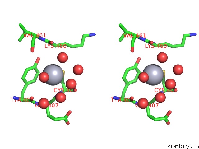
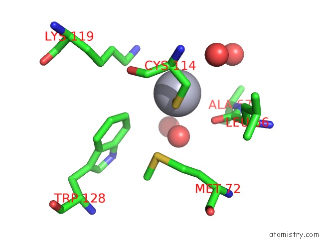
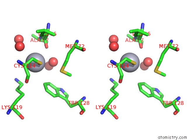
Mercury binding site 1 out of 3 in 1rwa
Go back to
Mercury binding site 1 out
of 3 in the Crystal Structure of Arthrobacter Aurescens Chondroitin Ac Lyase
Mono view
Stereo pair view
Mono view
Stereo pair view
A full contact list of Mercury with other atoms in the Hg binding
site number 1 of Crystal Structure of Arthrobacter Aurescens Chondroitin Ac Lyase within 5.0Å range:
|
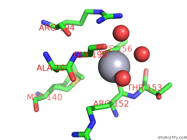
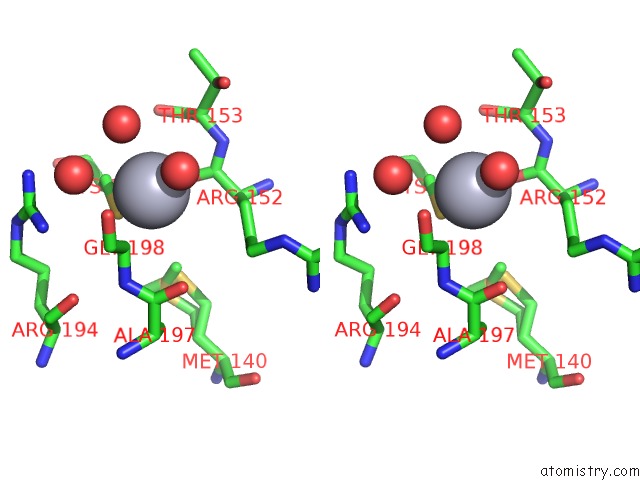
Mercury binding site 2 out of 3 in 1rwa
Go back to
Mercury binding site 2 out
of 3 in the Crystal Structure of Arthrobacter Aurescens Chondroitin Ac Lyase
Mono view
Stereo pair view
Mono view
Stereo pair view
A full contact list of Mercury with other atoms in the Hg binding
site number 2 of Crystal Structure of Arthrobacter Aurescens Chondroitin Ac Lyase within 5.0Å range:
|
Mercury binding site 3 out of 3 in 1rwa
Go back to
Mercury binding site 3 out
of 3 in the Crystal Structure of Arthrobacter Aurescens Chondroitin Ac Lyase
Mono view
Stereo pair view
Mono view
Stereo pair view
A full contact list of Mercury with other atoms in the Hg binding
site number 3 of Crystal Structure of Arthrobacter Aurescens Chondroitin Ac Lyase within 5.0Å range:
|
Reference:
V.V.Lunin,
Y.Li,
R.J.Linhardt,
H.Miyazono,
M.Kyogashima,
T.Kaneko,
A.W.Bell,
M.Cygler.
High-Resolution Crystal Structure of Arthrobacter Aurescens Chondroitin Ac Lyase: An Enzyme-Substrate Complex Defines the Catalytic Mechanism J.Mol.Biol. V. 337 367 2004.
ISSN: ISSN 0022-2836
PubMed: 15003453
DOI: 10.1016/J.JMB.2003.12.071
Page generated: Fri Aug 8 09:32:42 2025
ISSN: ISSN 0022-2836
PubMed: 15003453
DOI: 10.1016/J.JMB.2003.12.071
Last articles
K in 1YJ9K in 1YJ3
K in 1YIJ
K in 1YI2
K in 1YIT
K in 1YHQ
K in 1Y8P
K in 1Y8O
K in 1Y3S
K in 1Y39