Mercury »
PDB 1rsv-1yp2 »
1sms »
Mercury in PDB 1sms: Structure of the Ribonucleotide Reductase RNR4 Homodimer From Saccharomyces Cerevisiae
Enzymatic activity of Structure of the Ribonucleotide Reductase RNR4 Homodimer From Saccharomyces Cerevisiae
All present enzymatic activity of Structure of the Ribonucleotide Reductase RNR4 Homodimer From Saccharomyces Cerevisiae:
1.17.4.1;
1.17.4.1;
Protein crystallography data
The structure of Structure of the Ribonucleotide Reductase RNR4 Homodimer From Saccharomyces Cerevisiae, PDB code: 1sms
was solved by
M.Sommerhalter,
W.C.Voegtli,
D.L.Perlstein,
J.Ge,
J.Stubbe,
A.C.Rosenzweig,
with X-Ray Crystallography technique. A brief refinement statistics is given in the table below:
| Resolution Low / High (Å) | 12.00 / 3.10 |
| Space group | P 61 |
| Cell size a, b, c (Å), α, β, γ (°) | 79.600, 79.600, 218.100, 90.00, 90.00, 120.00 |
| R / Rfree (%) | 26.6 / 30.5 |
Mercury Binding Sites:
The binding sites of Mercury atom in the Structure of the Ribonucleotide Reductase RNR4 Homodimer From Saccharomyces Cerevisiae
(pdb code 1sms). This binding sites where shown within
5.0 Angstroms radius around Mercury atom.
In total 4 binding sites of Mercury where determined in the Structure of the Ribonucleotide Reductase RNR4 Homodimer From Saccharomyces Cerevisiae, PDB code: 1sms:
Jump to Mercury binding site number: 1; 2; 3; 4;
In total 4 binding sites of Mercury where determined in the Structure of the Ribonucleotide Reductase RNR4 Homodimer From Saccharomyces Cerevisiae, PDB code: 1sms:
Jump to Mercury binding site number: 1; 2; 3; 4;
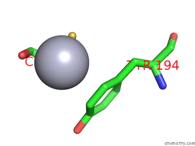
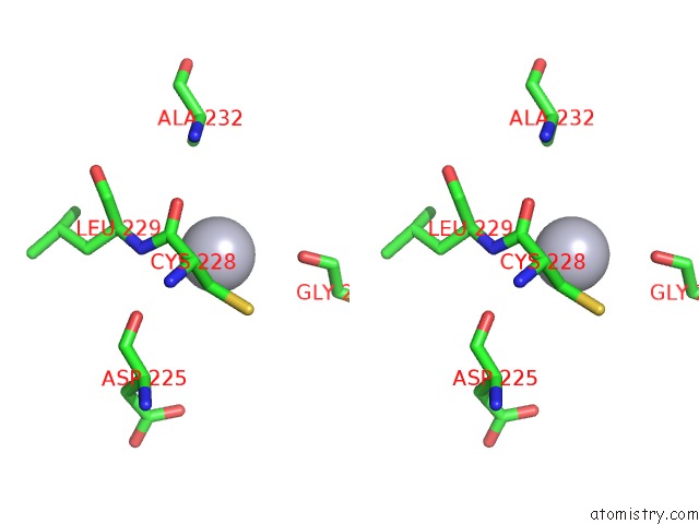
Mercury binding site 1 out of 4 in 1sms
Go back to
Mercury binding site 1 out
of 4 in the Structure of the Ribonucleotide Reductase RNR4 Homodimer From Saccharomyces Cerevisiae
Mono view
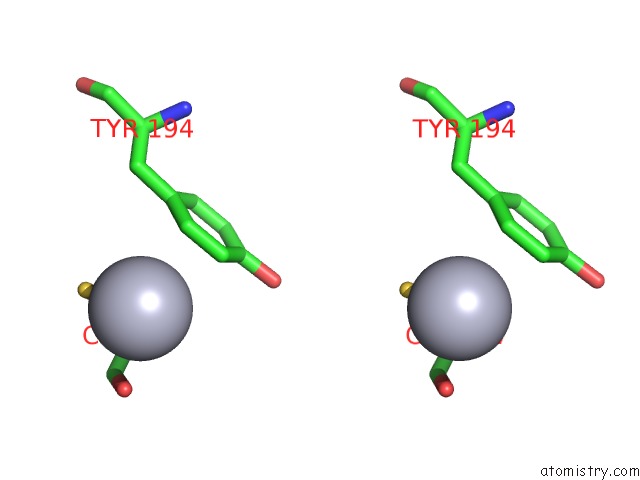
Stereo pair view
Mono view
Stereo pair view
A full contact list of Mercury with other atoms in the Hg binding
site number 1 of Structure of the Ribonucleotide Reductase RNR4 Homodimer From Saccharomyces Cerevisiae within 5.0Å range:
|
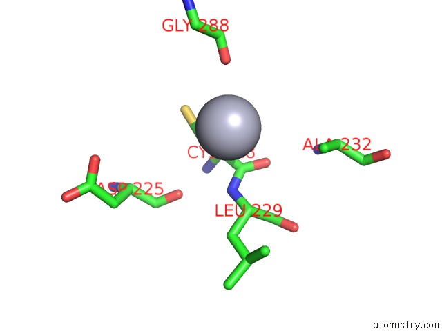
Mercury binding site 2 out of 4 in 1sms
Go back to
Mercury binding site 2 out
of 4 in the Structure of the Ribonucleotide Reductase RNR4 Homodimer From Saccharomyces Cerevisiae
Mono view

Stereo pair view
Mono view
Stereo pair view
A full contact list of Mercury with other atoms in the Hg binding
site number 2 of Structure of the Ribonucleotide Reductase RNR4 Homodimer From Saccharomyces Cerevisiae within 5.0Å range:
|
Mercury binding site 3 out of 4 in 1sms
Go back to
Mercury binding site 3 out
of 4 in the Structure of the Ribonucleotide Reductase RNR4 Homodimer From Saccharomyces Cerevisiae

Mono view
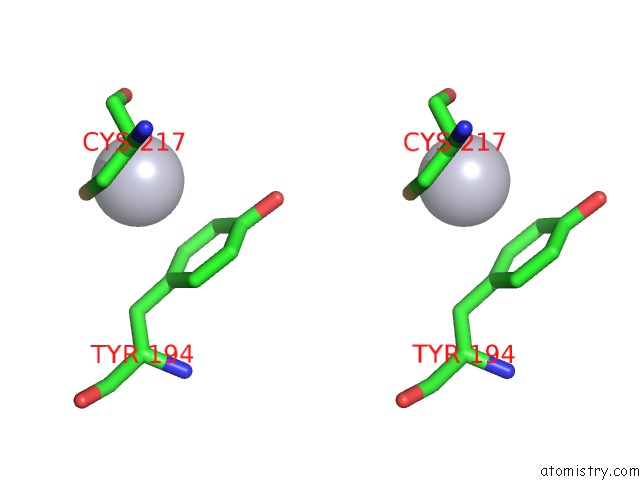
Stereo pair view
Mono view
Stereo pair view
A full contact list of Mercury with other atoms in the Hg binding
site number 3 of Structure of the Ribonucleotide Reductase RNR4 Homodimer From Saccharomyces Cerevisiae within 5.0Å range:
|
Mercury binding site 4 out of 4 in 1sms
Go back to
Mercury binding site 4 out
of 4 in the Structure of the Ribonucleotide Reductase RNR4 Homodimer From Saccharomyces Cerevisiae

Mono view
Stereo pair view
Mono view
Stereo pair view
A full contact list of Mercury with other atoms in the Hg binding
site number 4 of Structure of the Ribonucleotide Reductase RNR4 Homodimer From Saccharomyces Cerevisiae within 5.0Å range:
|
Reference:
M.Sommerhalter,
W.C.Voegtli,
D.L.Perlstein,
J.Ge,
J.Stubbe,
A.C.Rosenzweig.
Structures of the Yeast Ribonucleotide Reductase RNR2 and RNR4 Homodimers. Biochemistry V. 43 7736 2004.
ISSN: ISSN 0006-2960
PubMed: 15196016
DOI: 10.1021/BI049510M
Page generated: Fri Aug 8 09:32:52 2025
ISSN: ISSN 0006-2960
PubMed: 15196016
DOI: 10.1021/BI049510M
Last articles
I in 2GJMI in 2GSQ
I in 2GGQ
I in 2FWZ
I in 2G19
I in 2G1M
I in 2DGM
I in 2FWE
I in 2FWF
I in 2FWH