Mercury »
PDB 1rsv-1yp2 »
1yc9 »
Mercury in PDB 1yc9: The Crystal Structure of the Outer Membrane Protein Vcec From the Bacterial Pathogen Vibrio Cholerae at 1.8 Resolution
Protein crystallography data
The structure of The Crystal Structure of the Outer Membrane Protein Vcec From the Bacterial Pathogen Vibrio Cholerae at 1.8 Resolution, PDB code: 1yc9
was solved by
L.Federici,
D.Du,
F.Walas,
H.Matsumura,
J.Fernandez-Recio,
K.S.Mckeegan,
M.I.Borges-Walmsley,
B.F.Luisi,
A.R.Walmsley,
with X-Ray Crystallography technique. A brief refinement statistics is given in the table below:
| Resolution Low / High (Å) | 63.60 / 1.80 |
| Space group | P 3 2 1 |
| Cell size a, b, c (Å), α, β, γ (°) | 71.458, 71.458, 190.702, 90.00, 90.00, 120.00 |
| R / Rfree (%) | 18.9 / 22.1 |
Mercury Binding Sites:
The binding sites of Mercury atom in the The Crystal Structure of the Outer Membrane Protein Vcec From the Bacterial Pathogen Vibrio Cholerae at 1.8 Resolution
(pdb code 1yc9). This binding sites where shown within
5.0 Angstroms radius around Mercury atom.
In total 2 binding sites of Mercury where determined in the The Crystal Structure of the Outer Membrane Protein Vcec From the Bacterial Pathogen Vibrio Cholerae at 1.8 Resolution, PDB code: 1yc9:
Jump to Mercury binding site number: 1; 2;
In total 2 binding sites of Mercury where determined in the The Crystal Structure of the Outer Membrane Protein Vcec From the Bacterial Pathogen Vibrio Cholerae at 1.8 Resolution, PDB code: 1yc9:
Jump to Mercury binding site number: 1; 2;
Mercury binding site 1 out of 2 in 1yc9
Go back to
Mercury binding site 1 out
of 2 in the The Crystal Structure of the Outer Membrane Protein Vcec From the Bacterial Pathogen Vibrio Cholerae at 1.8 Resolution
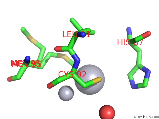
Mono view
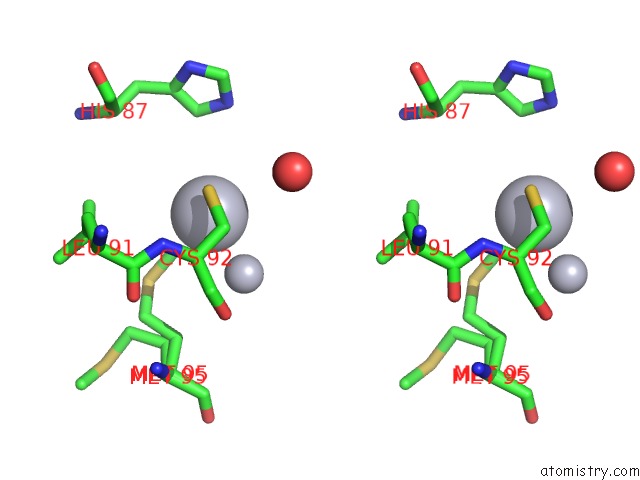
Stereo pair view
Mono view
Stereo pair view
A full contact list of Mercury with other atoms in the Hg binding
site number 1 of The Crystal Structure of the Outer Membrane Protein Vcec From the Bacterial Pathogen Vibrio Cholerae at 1.8 Resolution within 5.0Å range:
|
Mercury binding site 2 out of 2 in 1yc9
Go back to
Mercury binding site 2 out
of 2 in the The Crystal Structure of the Outer Membrane Protein Vcec From the Bacterial Pathogen Vibrio Cholerae at 1.8 Resolution
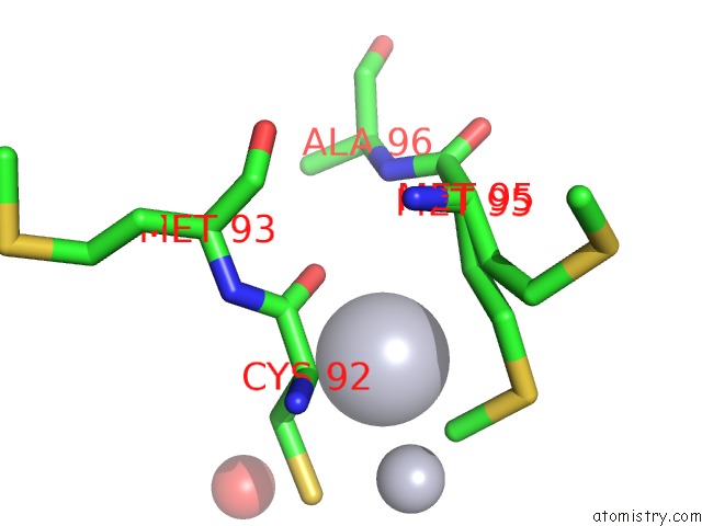
Mono view
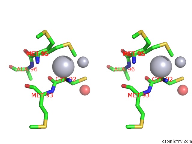
Stereo pair view
Mono view
Stereo pair view
A full contact list of Mercury with other atoms in the Hg binding
site number 2 of The Crystal Structure of the Outer Membrane Protein Vcec From the Bacterial Pathogen Vibrio Cholerae at 1.8 Resolution within 5.0Å range:
|
Reference:
L.Federici,
D.Du,
F.Walas,
H.Matsumura,
J.Fernandez-Recio,
K.S.Mckeegan,
M.I.Borges-Walmsley,
B.F.Luisi,
A.R.Walmsley.
The Crystal Structure of the Outer Membrane Protein Vcec From the Bacterial Pathogen Vibrio Cholerae at 1.8 A Resolution J.Biol.Chem. V. 280 15307 2005.
ISSN: ISSN 0021-9258
PubMed: 15684414
DOI: 10.1074/JBC.M500401200
Page generated: Fri Aug 8 09:38:52 2025
ISSN: ISSN 0021-9258
PubMed: 15684414
DOI: 10.1074/JBC.M500401200
Last articles
K in 2ANNK in 2AL1
K in 2AL2
K in 2AJ7
K in 2ADQ
K in 2ADP
K in 2A79
K in 2A2O
K in 2AAQ
K in 2A6X