Mercury »
PDB 1yu1-2epm »
2alx »
Mercury in PDB 2alx: Ribonucleotide Reductase R2 From Escherichia Coli in Space Group P6(1)22
Enzymatic activity of Ribonucleotide Reductase R2 From Escherichia Coli in Space Group P6(1)22
All present enzymatic activity of Ribonucleotide Reductase R2 From Escherichia Coli in Space Group P6(1)22:
1.17.4.1;
1.17.4.1;
Protein crystallography data
The structure of Ribonucleotide Reductase R2 From Escherichia Coli in Space Group P6(1)22, PDB code: 2alx
was solved by
M.Sommerhalter,
L.Saleh,
J.M.Bollinger Jr.,
A.C.Rosenzweig,
with X-Ray Crystallography technique. A brief refinement statistics is given in the table below:
| Resolution Low / High (Å) | 19.27 / 2.60 |
| Space group | P 61 2 2 |
| Cell size a, b, c (Å), α, β, γ (°) | 92.940, 92.940, 200.930, 90.00, 90.00, 120.00 |
| R / Rfree (%) | 22.7 / 27.6 |
Other elements in 2alx:
The structure of Ribonucleotide Reductase R2 From Escherichia Coli in Space Group P6(1)22 also contains other interesting chemical elements:
| Manganese | (Mn) | 2 atoms |
Mercury Binding Sites:
The binding sites of Mercury atom in the Ribonucleotide Reductase R2 From Escherichia Coli in Space Group P6(1)22
(pdb code 2alx). This binding sites where shown within
5.0 Angstroms radius around Mercury atom.
In total 4 binding sites of Mercury where determined in the Ribonucleotide Reductase R2 From Escherichia Coli in Space Group P6(1)22, PDB code: 2alx:
Jump to Mercury binding site number: 1; 2; 3; 4;
In total 4 binding sites of Mercury where determined in the Ribonucleotide Reductase R2 From Escherichia Coli in Space Group P6(1)22, PDB code: 2alx:
Jump to Mercury binding site number: 1; 2; 3; 4;
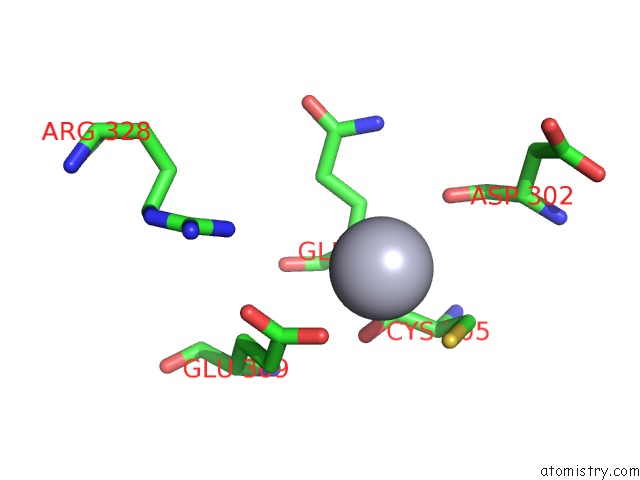
Mercury binding site 1 out of 4 in 2alx
Go back to
Mercury binding site 1 out
of 4 in the Ribonucleotide Reductase R2 From Escherichia Coli in Space Group P6(1)22
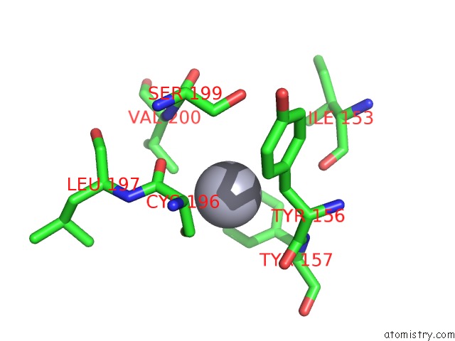
Mono view
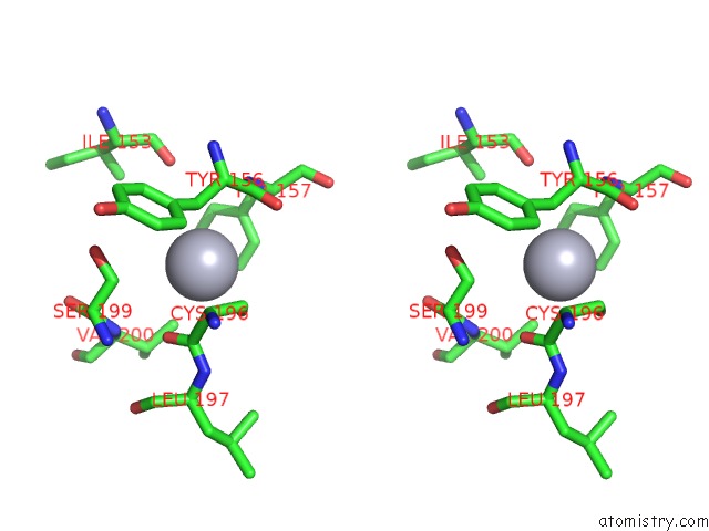
Stereo pair view
Mono view
Stereo pair view
A full contact list of Mercury with other atoms in the Hg binding
site number 1 of Ribonucleotide Reductase R2 From Escherichia Coli in Space Group P6(1)22 within 5.0Å range:
|
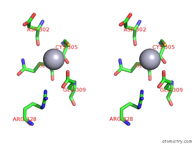
Mercury binding site 2 out of 4 in 2alx
Go back to
Mercury binding site 2 out
of 4 in the Ribonucleotide Reductase R2 From Escherichia Coli in Space Group P6(1)22
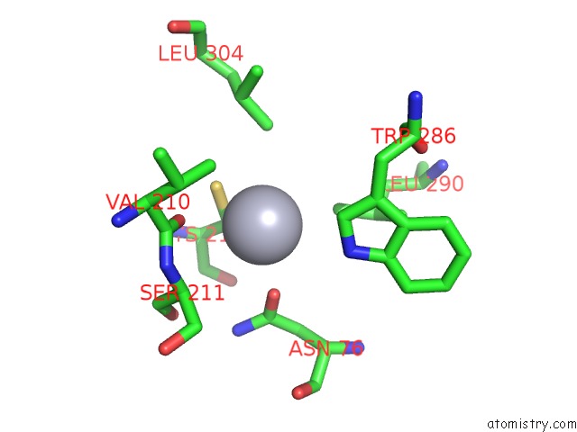
Mono view
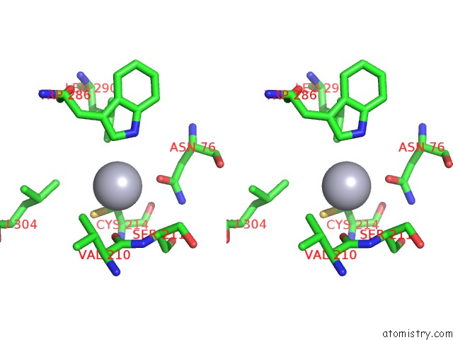
Stereo pair view
Mono view
Stereo pair view
A full contact list of Mercury with other atoms in the Hg binding
site number 2 of Ribonucleotide Reductase R2 From Escherichia Coli in Space Group P6(1)22 within 5.0Å range:
|
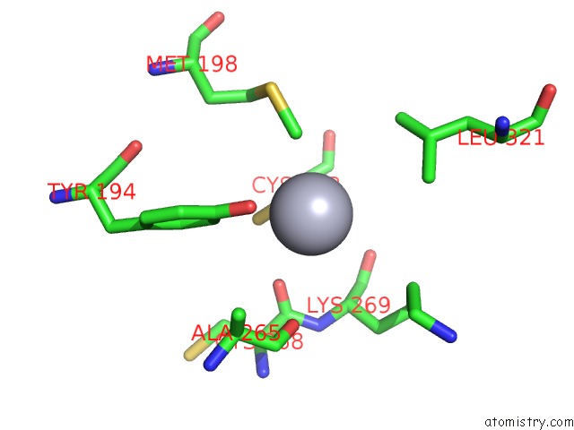
Mercury binding site 3 out of 4 in 2alx
Go back to
Mercury binding site 3 out
of 4 in the Ribonucleotide Reductase R2 From Escherichia Coli in Space Group P6(1)22
Mono view
Stereo pair view
Mono view
Stereo pair view
A full contact list of Mercury with other atoms in the Hg binding
site number 3 of Ribonucleotide Reductase R2 From Escherichia Coli in Space Group P6(1)22 within 5.0Å range:
|
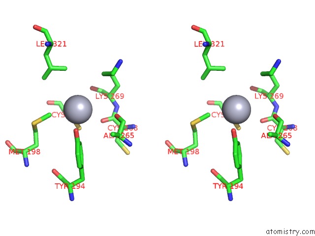
Mercury binding site 4 out of 4 in 2alx
Go back to
Mercury binding site 4 out
of 4 in the Ribonucleotide Reductase R2 From Escherichia Coli in Space Group P6(1)22
Mono view
Stereo pair view
Mono view
Stereo pair view
A full contact list of Mercury with other atoms in the Hg binding
site number 4 of Ribonucleotide Reductase R2 From Escherichia Coli in Space Group P6(1)22 within 5.0Å range:
|
Reference:
M.Sommerhalter,
L.Saleh,
J.M.Bollinger,
A.C.Rosenzweig.
Structure of Escherichia Coli Ribonucleotide Reductase R2 in Space Group P6122. Acta Crystallogr.,Sect.D V. 61 1649 2005.
ISSN: ISSN 0907-4449
PubMed: 16301799
DOI: 10.1107/S0907444905034062
Page generated: Fri Aug 8 09:45:22 2025
ISSN: ISSN 0907-4449
PubMed: 16301799
DOI: 10.1107/S0907444905034062
Last articles
I in 4PVGI in 4PGC
I in 4PNS
I in 4P4Z
I in 4P1E
I in 4P4X
I in 4P4Y
I in 4P4W
I in 4OA5
I in 4OWA