Mercury »
PDB 1yu1-2epm »
2dpm »
Mercury in PDB 2dpm: Dpnm Dna Adenine Methyltransferase From Streptoccocus Pneumoniae Complexed with S-Adenosylmethionine
Enzymatic activity of Dpnm Dna Adenine Methyltransferase From Streptoccocus Pneumoniae Complexed with S-Adenosylmethionine
All present enzymatic activity of Dpnm Dna Adenine Methyltransferase From Streptoccocus Pneumoniae Complexed with S-Adenosylmethionine:
2.1.1.72;
2.1.1.72;
Protein crystallography data
The structure of Dpnm Dna Adenine Methyltransferase From Streptoccocus Pneumoniae Complexed with S-Adenosylmethionine, PDB code: 2dpm
was solved by
P.H.Tran,
Z.R.Korszun,
S.Cerritelli,
S.S.Springhorn,
S.A.Lacks,
with X-Ray Crystallography technique. A brief refinement statistics is given in the table below:
| Resolution Low / High (Å) | 20.00 / 1.80 |
| Space group | P 21 21 21 |
| Cell size a, b, c (Å), α, β, γ (°) | 56.600, 68.100, 85.200, 90.00, 90.00, 90.00 |
| R / Rfree (%) | 23.8 / 28.4 |
Mercury Binding Sites:
The binding sites of Mercury atom in the Dpnm Dna Adenine Methyltransferase From Streptoccocus Pneumoniae Complexed with S-Adenosylmethionine
(pdb code 2dpm). This binding sites where shown within
5.0 Angstroms radius around Mercury atom.
In total only one binding site of Mercury was determined in the Dpnm Dna Adenine Methyltransferase From Streptoccocus Pneumoniae Complexed with S-Adenosylmethionine, PDB code: 2dpm:
In total only one binding site of Mercury was determined in the Dpnm Dna Adenine Methyltransferase From Streptoccocus Pneumoniae Complexed with S-Adenosylmethionine, PDB code: 2dpm:
Mercury binding site 1 out of 1 in 2dpm
Go back to
Mercury binding site 1 out
of 1 in the Dpnm Dna Adenine Methyltransferase From Streptoccocus Pneumoniae Complexed with S-Adenosylmethionine
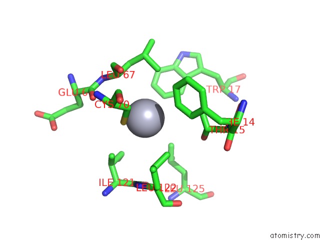
Mono view
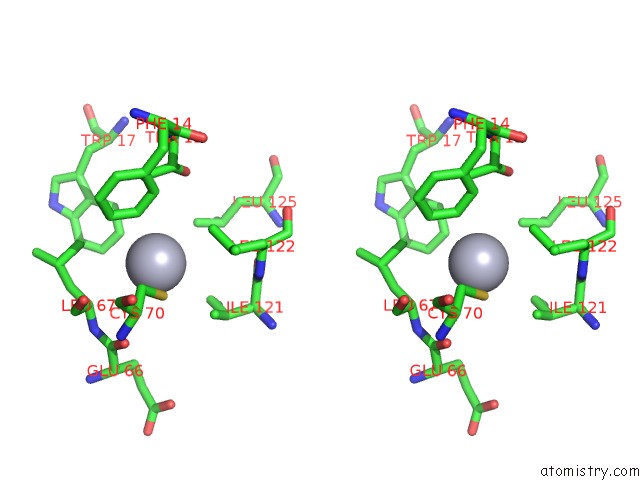
Stereo pair view
Mono view
Stereo pair view
A full contact list of Mercury with other atoms in the Hg binding
site number 1 of Dpnm Dna Adenine Methyltransferase From Streptoccocus Pneumoniae Complexed with S-Adenosylmethionine within 5.0Å range:
|
Reference:
P.H.Tran,
Z.R.Korszun,
S.Cerritelli,
S.S.Springhorn,
S.A.Lacks.
Crystal Structure of the Dpnm Dna Adenine Methyltransferase From the Dpnii Restriction System of Streptococcus Pneumoniae Bound to S-Adenosylmethionine. Structure V. 6 1563 1998.
ISSN: ISSN 0969-2126
PubMed: 9862809
DOI: 10.1016/S0969-2126(98)00154-3
Page generated: Sun Aug 11 02:25:32 2024
ISSN: ISSN 0969-2126
PubMed: 9862809
DOI: 10.1016/S0969-2126(98)00154-3
Last articles
Cl in 2RABCl in 2RA6
Cl in 2R9Z
Cl in 2R9H
Cl in 2R9V
Cl in 2R9F
Cl in 2R9K
Cl in 2R9C
Cl in 2R83
Cl in 2R8X