Mercury »
PDB 2esw-2o1f »
2f14 »
Mercury in PDB 2f14: Tne Crystal Structure of the Human Carbonic Anhydrase II in Complex with A Fluorescent Inhibitor
Enzymatic activity of Tne Crystal Structure of the Human Carbonic Anhydrase II in Complex with A Fluorescent Inhibitor
All present enzymatic activity of Tne Crystal Structure of the Human Carbonic Anhydrase II in Complex with A Fluorescent Inhibitor:
4.2.1.1;
4.2.1.1;
Protein crystallography data
The structure of Tne Crystal Structure of the Human Carbonic Anhydrase II in Complex with A Fluorescent Inhibitor, PDB code: 2f14
was solved by
V.Alterio,
C.Pedone,
G.De Simone,
with X-Ray Crystallography technique. A brief refinement statistics is given in the table below:
| Resolution Low / High (Å) | 20.00 / 1.71 |
| Space group | P 1 21 1 |
| Cell size a, b, c (Å), α, β, γ (°) | 42.260, 41.600, 72.220, 90.00, 104.57, 90.00 |
| R / Rfree (%) | 17.2 / 20.2 |
Other elements in 2f14:
The structure of Tne Crystal Structure of the Human Carbonic Anhydrase II in Complex with A Fluorescent Inhibitor also contains other interesting chemical elements:
| Zinc | (Zn) | 1 atom |
Mercury Binding Sites:
The binding sites of Mercury atom in the Tne Crystal Structure of the Human Carbonic Anhydrase II in Complex with A Fluorescent Inhibitor
(pdb code 2f14). This binding sites where shown within
5.0 Angstroms radius around Mercury atom.
In total only one binding site of Mercury was determined in the Tne Crystal Structure of the Human Carbonic Anhydrase II in Complex with A Fluorescent Inhibitor, PDB code: 2f14:
In total only one binding site of Mercury was determined in the Tne Crystal Structure of the Human Carbonic Anhydrase II in Complex with A Fluorescent Inhibitor, PDB code: 2f14:
Mercury binding site 1 out of 1 in 2f14
Go back to
Mercury binding site 1 out
of 1 in the Tne Crystal Structure of the Human Carbonic Anhydrase II in Complex with A Fluorescent Inhibitor
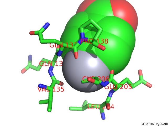
Mono view
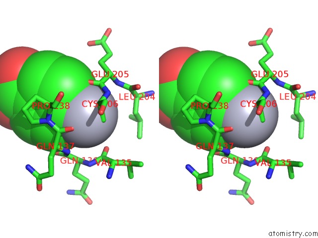
Stereo pair view
Mono view
Stereo pair view
A full contact list of Mercury with other atoms in the Hg binding
site number 1 of Tne Crystal Structure of the Human Carbonic Anhydrase II in Complex with A Fluorescent Inhibitor within 5.0Å range:
|
Reference:
V.Alterio,
R.M.Vitale,
S.M.Monti,
C.Pedone,
A.Scozzafava,
A.Cecchi,
G.De Simone,
C.T.Supuran.
Carbonic Anhydrase Inhibitors: X-Ray and Molecular Modeling Study For the Interaction of A Fluorescent Antitumor Sulfonamide with Isozyme II and IX. J.Am.Chem.Soc. V. 128 8329 2006.
ISSN: ISSN 0002-7863
PubMed: 16787097
DOI: 10.1021/JA061574S
Page generated: Sun Aug 11 02:33:12 2024
ISSN: ISSN 0002-7863
PubMed: 16787097
DOI: 10.1021/JA061574S
Last articles
Zn in 9MJ5Zn in 9HNW
Zn in 9G0L
Zn in 9FNE
Zn in 9DZN
Zn in 9E0I
Zn in 9D32
Zn in 9DAK
Zn in 8ZXC
Zn in 8ZUF