Mercury »
PDB 2esw-2o1f »
2fxm »
Mercury in PDB 2fxm: Structure of the Human Beta-Myosin S2 Fragment
Protein crystallography data
The structure of Structure of the Human Beta-Myosin S2 Fragment, PDB code: 2fxm
was solved by
W.Blankenfeldt,
N.H.Thoma,
J.S.Wray,
M.Gautel,
I.Schlichting,
with X-Ray Crystallography technique. A brief refinement statistics is given in the table below:
| Resolution Low / High (Å) | 20.00 / 2.70 |
| Space group | C 2 2 21 |
| Cell size a, b, c (Å), α, β, γ (°) | 40.290, 45.970, 373.700, 90.00, 90.00, 90.00 |
| R / Rfree (%) | 24.2 / 28.3 |
Mercury Binding Sites:
The binding sites of Mercury atom in the Structure of the Human Beta-Myosin S2 Fragment
(pdb code 2fxm). This binding sites where shown within
5.0 Angstroms radius around Mercury atom.
In total 2 binding sites of Mercury where determined in the Structure of the Human Beta-Myosin S2 Fragment, PDB code: 2fxm:
Jump to Mercury binding site number: 1; 2;
In total 2 binding sites of Mercury where determined in the Structure of the Human Beta-Myosin S2 Fragment, PDB code: 2fxm:
Jump to Mercury binding site number: 1; 2;
Mercury binding site 1 out of 2 in 2fxm
Go back to
Mercury binding site 1 out
of 2 in the Structure of the Human Beta-Myosin S2 Fragment
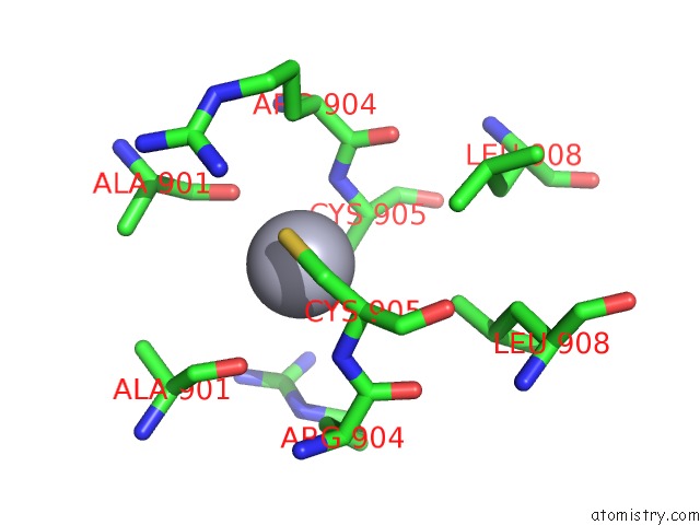
Mono view
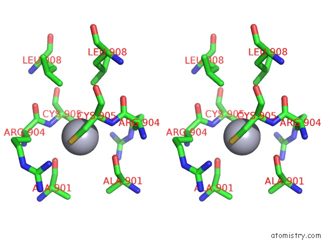
Stereo pair view
Mono view
Stereo pair view
A full contact list of Mercury with other atoms in the Hg binding
site number 1 of Structure of the Human Beta-Myosin S2 Fragment within 5.0Å range:
|
Mercury binding site 2 out of 2 in 2fxm
Go back to
Mercury binding site 2 out
of 2 in the Structure of the Human Beta-Myosin S2 Fragment
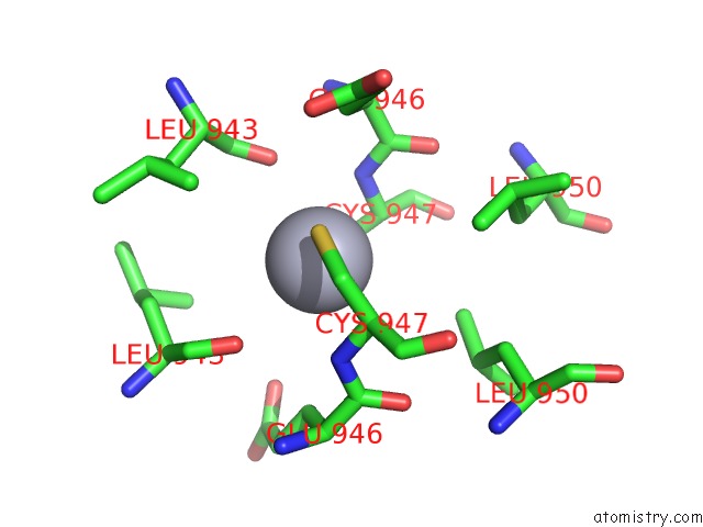
Mono view
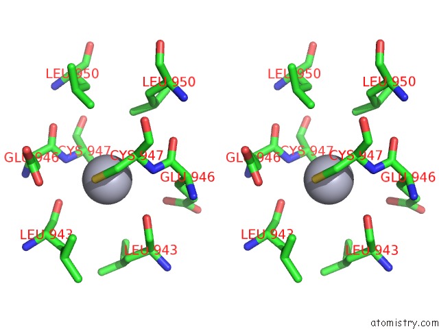
Stereo pair view
Mono view
Stereo pair view
A full contact list of Mercury with other atoms in the Hg binding
site number 2 of Structure of the Human Beta-Myosin S2 Fragment within 5.0Å range:
|
Reference:
W.Blankenfeldt,
N.H.Thoma,
J.S.Wray,
M.Gautel,
I.Schlichting.
Crystal Structures of Human Cardiac {Beta}-Myosin II S2-{Delta} Provide Insight Into the Functional Role of the S2 Subfragment Proc.Natl.Acad.Sci.Usa V. 103 17713 2006.
ISSN: ISSN 0027-8424
PubMed: 17095604
DOI: 10.1073/PNAS.0606741103
Page generated: Sun Aug 11 02:34:57 2024
ISSN: ISSN 0027-8424
PubMed: 17095604
DOI: 10.1073/PNAS.0606741103
Last articles
Zn in 9J0NZn in 9J0O
Zn in 9J0P
Zn in 9FJX
Zn in 9EKB
Zn in 9C0F
Zn in 9CAH
Zn in 9CH0
Zn in 9CH3
Zn in 9CH1