Mercury »
PDB 2esw-2o1f »
2j0e »
Mercury in PDB 2j0e: Three Dimensional Structure and Catalytic Mechanism of 6- Phosphogluconolactonase From Trypanosoma Brucei
Enzymatic activity of Three Dimensional Structure and Catalytic Mechanism of 6- Phosphogluconolactonase From Trypanosoma Brucei
All present enzymatic activity of Three Dimensional Structure and Catalytic Mechanism of 6- Phosphogluconolactonase From Trypanosoma Brucei:
3.1.1.31;
3.1.1.31;
Protein crystallography data
The structure of Three Dimensional Structure and Catalytic Mechanism of 6- Phosphogluconolactonase From Trypanosoma Brucei, PDB code: 2j0e
was solved by
M.Delarue,
N.Duclert-Savatier,
E.Miclet,
A.Haouz,
D.Giganti,
J.Ouazzani,
P.Lopez,
M.Nilges,
V.Stoven,
with X-Ray Crystallography technique. A brief refinement statistics is given in the table below:
| Resolution Low / High (Å) | 18.00 / 2.1 |
| Space group | P 21 21 21 |
| Cell size a, b, c (Å), α, β, γ (°) | 70.310, 80.850, 90.310, 90.00, 90.00, 90.00 |
| R / Rfree (%) | 20.6 / 24.6 |
Other elements in 2j0e:
The structure of Three Dimensional Structure and Catalytic Mechanism of 6- Phosphogluconolactonase From Trypanosoma Brucei also contains other interesting chemical elements:
| Potassium | (K) | 1 atom |
| Zinc | (Zn) | 2 atoms |
Mercury Binding Sites:
The binding sites of Mercury atom in the Three Dimensional Structure and Catalytic Mechanism of 6- Phosphogluconolactonase From Trypanosoma Brucei
(pdb code 2j0e). This binding sites where shown within
5.0 Angstroms radius around Mercury atom.
In total 4 binding sites of Mercury where determined in the Three Dimensional Structure and Catalytic Mechanism of 6- Phosphogluconolactonase From Trypanosoma Brucei, PDB code: 2j0e:
Jump to Mercury binding site number: 1; 2; 3; 4;
In total 4 binding sites of Mercury where determined in the Three Dimensional Structure and Catalytic Mechanism of 6- Phosphogluconolactonase From Trypanosoma Brucei, PDB code: 2j0e:
Jump to Mercury binding site number: 1; 2; 3; 4;
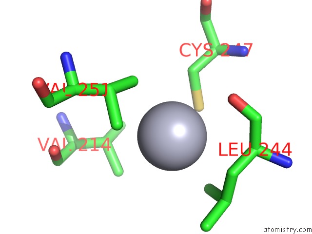
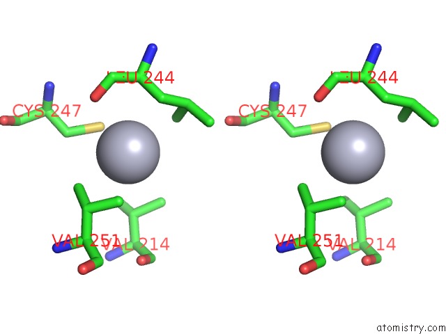
Mercury binding site 1 out of 4 in 2j0e
Go back to
Mercury binding site 1 out
of 4 in the Three Dimensional Structure and Catalytic Mechanism of 6- Phosphogluconolactonase From Trypanosoma Brucei
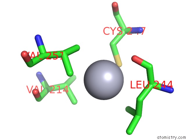
Mono view
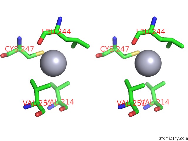
Stereo pair view
Mono view
Stereo pair view
A full contact list of Mercury with other atoms in the Hg binding
site number 1 of Three Dimensional Structure and Catalytic Mechanism of 6- Phosphogluconolactonase From Trypanosoma Brucei within 5.0Å range:
|
Mercury binding site 2 out of 4 in 2j0e
Go back to
Mercury binding site 2 out
of 4 in the Three Dimensional Structure and Catalytic Mechanism of 6- Phosphogluconolactonase From Trypanosoma Brucei
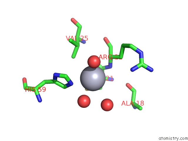
Mono view
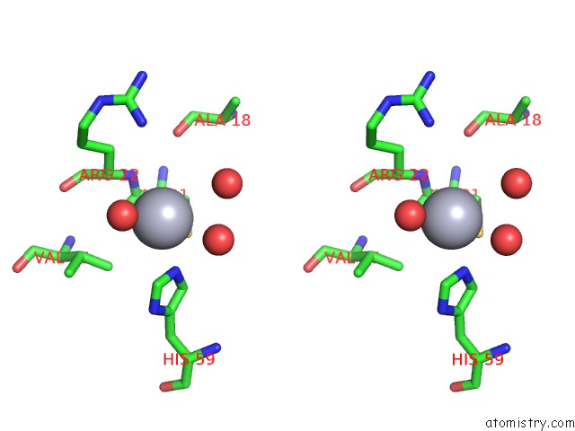
Stereo pair view
Mono view
Stereo pair view
A full contact list of Mercury with other atoms in the Hg binding
site number 2 of Three Dimensional Structure and Catalytic Mechanism of 6- Phosphogluconolactonase From Trypanosoma Brucei within 5.0Å range:
|
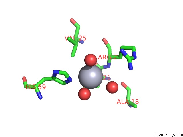
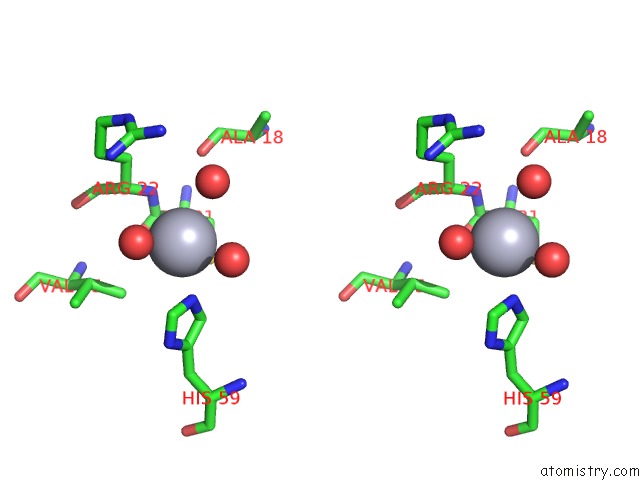
Mercury binding site 3 out of 4 in 2j0e
Go back to
Mercury binding site 3 out
of 4 in the Three Dimensional Structure and Catalytic Mechanism of 6- Phosphogluconolactonase From Trypanosoma Brucei
Mono view
Stereo pair view
Mono view
Stereo pair view
A full contact list of Mercury with other atoms in the Hg binding
site number 3 of Three Dimensional Structure and Catalytic Mechanism of 6- Phosphogluconolactonase From Trypanosoma Brucei within 5.0Å range:
|
Mercury binding site 4 out of 4 in 2j0e
Go back to
Mercury binding site 4 out
of 4 in the Three Dimensional Structure and Catalytic Mechanism of 6- Phosphogluconolactonase From Trypanosoma Brucei
Mono view
Stereo pair view
Mono view
Stereo pair view
A full contact list of Mercury with other atoms in the Hg binding
site number 4 of Three Dimensional Structure and Catalytic Mechanism of 6- Phosphogluconolactonase From Trypanosoma Brucei within 5.0Å range:
|
Reference:
M.Delarue,
N.Duclert-Savatier,
E.Miclet,
A.Haouz,
D.Giganti,
J.Ouazzani,
P.Lopez,
M.Nilges,
V.Stoven.
Three Dimensional Structure and Implications For the Catalytic Mechanism of 6- Phosphogluconolactonase From Trypanosoma Brucei. J.Mol.Biol. V. 366 868 2007.
ISSN: ISSN 0022-2836
PubMed: 17196981
DOI: 10.1016/J.JMB.2006.11.063
Page generated: Fri Aug 8 09:51:22 2025
ISSN: ISSN 0022-2836
PubMed: 17196981
DOI: 10.1016/J.JMB.2006.11.063
Last articles
I in 2EDAI in 2DFC
I in 2CJ7
I in 2D8P
I in 2D97
I in 2D8W
I in 2D91
I in 2D8O
I in 2CJ8
I in 2CKK