Mercury »
PDB 2o1g-3b4f »
2p4e »
Mercury in PDB 2p4e: Crystal Structure of PCSK9
Protein crystallography data
The structure of Crystal Structure of PCSK9, PDB code: 2p4e
was solved by
D.Cunningham,
D.E.Danley,
F.K.Geoghegan,
M.C.Griffor,
J.L.Hawkins,
X.Qiu,
with X-Ray Crystallography technique. A brief refinement statistics is given in the table below:
| Resolution Low / High (Å) | 75.00 / 1.98 |
| Space group | P 21 21 21 |
| Cell size a, b, c (Å), α, β, γ (°) | 62.810, 70.670, 150.020, 90.00, 90.00, 90.00 |
| R / Rfree (%) | 20 / 25 |
Mercury Binding Sites:
The binding sites of Mercury atom in the Crystal Structure of PCSK9
(pdb code 2p4e). This binding sites where shown within
5.0 Angstroms radius around Mercury atom.
In total 2 binding sites of Mercury where determined in the Crystal Structure of PCSK9, PDB code: 2p4e:
Jump to Mercury binding site number: 1; 2;
In total 2 binding sites of Mercury where determined in the Crystal Structure of PCSK9, PDB code: 2p4e:
Jump to Mercury binding site number: 1; 2;
Mercury binding site 1 out of 2 in 2p4e
Go back to
Mercury binding site 1 out
of 2 in the Crystal Structure of PCSK9
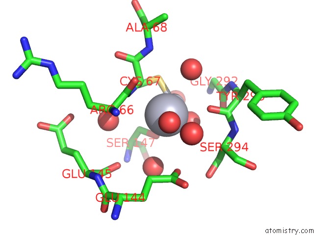
Mono view
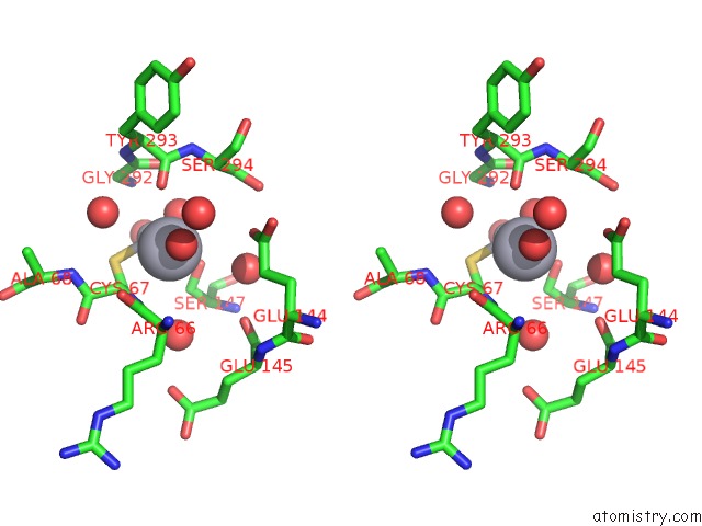
Stereo pair view
Mono view
Stereo pair view
A full contact list of Mercury with other atoms in the Hg binding
site number 1 of Crystal Structure of PCSK9 within 5.0Å range:
|
Mercury binding site 2 out of 2 in 2p4e
Go back to
Mercury binding site 2 out
of 2 in the Crystal Structure of PCSK9
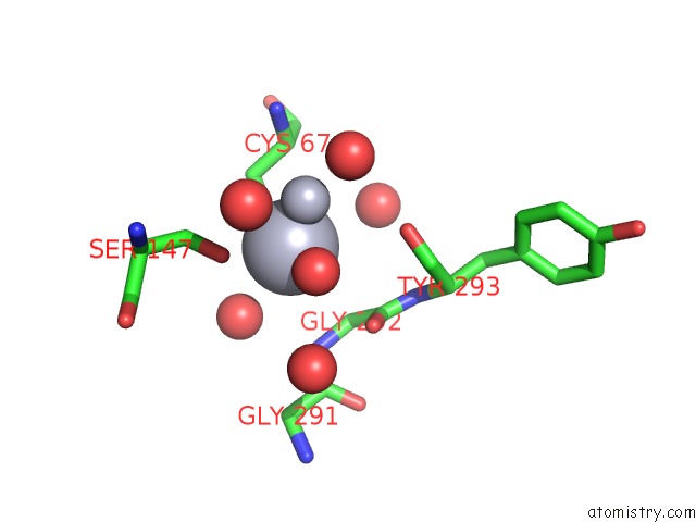
Mono view
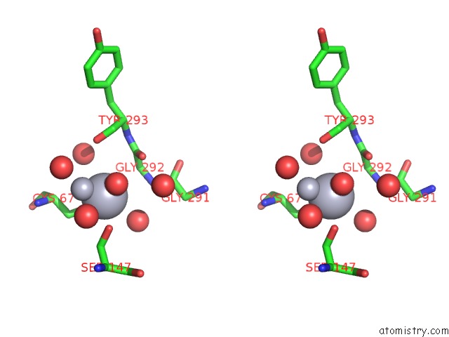
Stereo pair view
Mono view
Stereo pair view
A full contact list of Mercury with other atoms in the Hg binding
site number 2 of Crystal Structure of PCSK9 within 5.0Å range:
|
Reference:
D.Cunningham,
D.E.Danley,
K.F.Geoghegan,
M.C.Griffor,
J.L.Hawkins,
T.A.Subashi,
A.H.Varghese,
M.J.Ammirati,
J.S.Culp,
L.R.Hoth,
M.N.Mansour,
K.M.Mcgrath,
A.P.Seddon,
S.Shenolikar,
K.J.Stutzman-Engwall,
L.C.Warren,
D.Xia,
X.Qiu.
Structural and Biophysical Studies of PCSK9 and Its Mutants Linked to Familial Hypercholesterolemia. Nat.Struct.Mol.Biol. V. 14 413 2007.
ISSN: ISSN 1545-9993
PubMed: 17435765
DOI: 10.1038/NSMB1235
Page generated: Sun Aug 11 02:53:29 2024
ISSN: ISSN 1545-9993
PubMed: 17435765
DOI: 10.1038/NSMB1235
Last articles
Zn in 9MJ5Zn in 9HNW
Zn in 9G0L
Zn in 9FNE
Zn in 9DZN
Zn in 9E0I
Zn in 9D32
Zn in 9DAK
Zn in 8ZXC
Zn in 8ZUF