Mercury »
PDB 3bl0-3k7k »
3d8d »
Mercury in PDB 3d8d: Crystal Structure of the Human FE65-PTB1 Domain
Protein crystallography data
The structure of Crystal Structure of the Human FE65-PTB1 Domain, PDB code: 3d8d
was solved by
J.Radzimanowski,
S.Ravaud,
I.Sinning,
K.Wild,
with X-Ray Crystallography technique. A brief refinement statistics is given in the table below:
| Resolution Low / High (Å) | 42.00 / 2.20 |
| Space group | P 21 21 21 |
| Cell size a, b, c (Å), α, β, γ (°) | 43.756, 79.574, 83.974, 90.00, 90.00, 90.00 |
| R / Rfree (%) | 20.2 / 25.2 |
Mercury Binding Sites:
The binding sites of Mercury atom in the Crystal Structure of the Human FE65-PTB1 Domain
(pdb code 3d8d). This binding sites where shown within
5.0 Angstroms radius around Mercury atom.
In total 6 binding sites of Mercury where determined in the Crystal Structure of the Human FE65-PTB1 Domain, PDB code: 3d8d:
Jump to Mercury binding site number: 1; 2; 3; 4; 5; 6;
In total 6 binding sites of Mercury where determined in the Crystal Structure of the Human FE65-PTB1 Domain, PDB code: 3d8d:
Jump to Mercury binding site number: 1; 2; 3; 4; 5; 6;
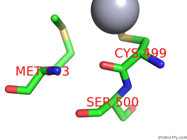
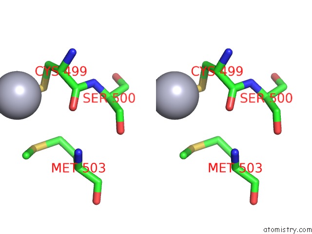
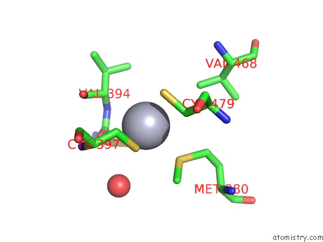
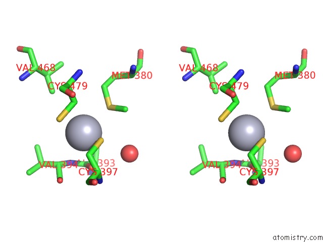
Mercury binding site 1 out of 6 in 3d8d
Go back to
Mercury binding site 1 out
of 6 in the Crystal Structure of the Human FE65-PTB1 Domain
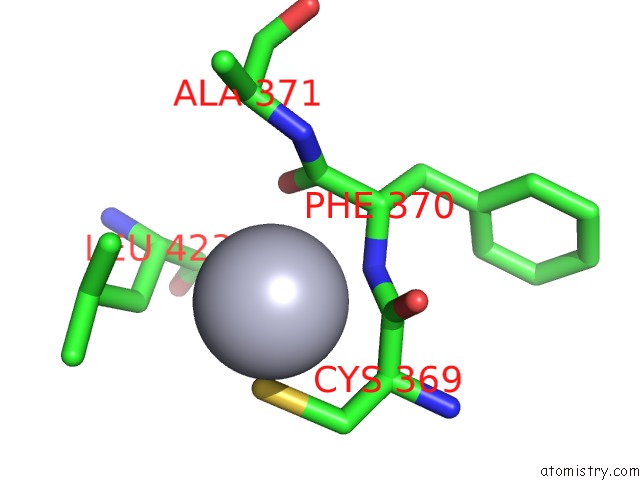
Mono view
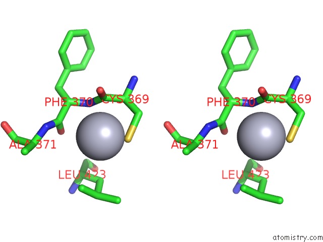
Stereo pair view
Mono view
Stereo pair view
A full contact list of Mercury with other atoms in the Hg binding
site number 1 of Crystal Structure of the Human FE65-PTB1 Domain within 5.0Å range:
|
Mercury binding site 2 out of 6 in 3d8d
Go back to
Mercury binding site 2 out
of 6 in the Crystal Structure of the Human FE65-PTB1 Domain
Mono view
Stereo pair view
Mono view
Stereo pair view
A full contact list of Mercury with other atoms in the Hg binding
site number 2 of Crystal Structure of the Human FE65-PTB1 Domain within 5.0Å range:
|
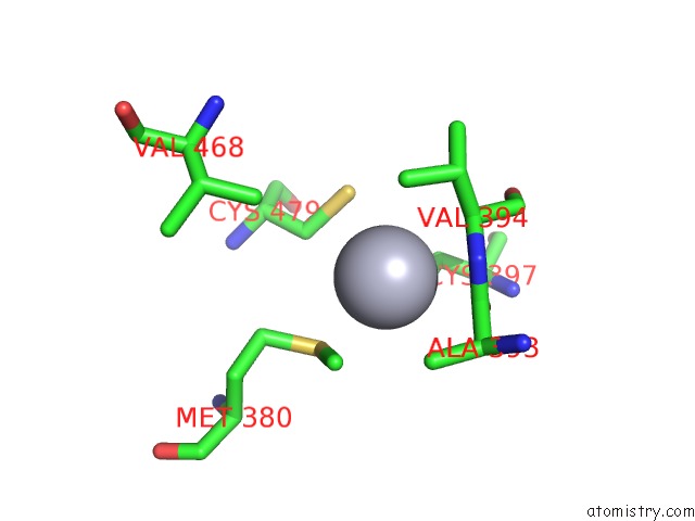
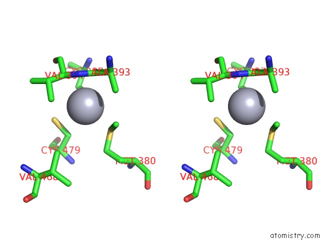
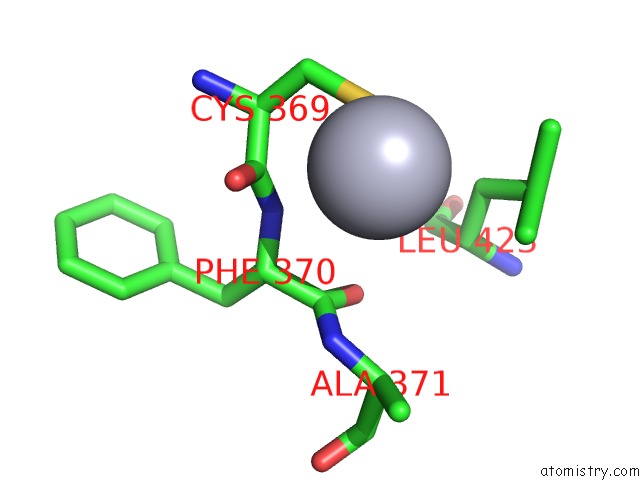
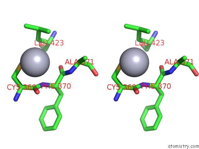
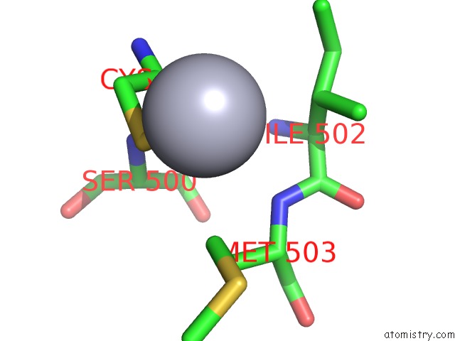
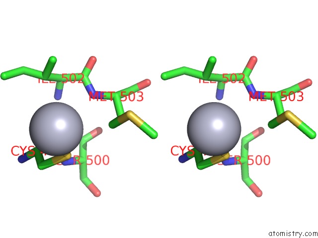
Mercury binding site 3 out of 6 in 3d8d
Go back to
Mercury binding site 3 out
of 6 in the Crystal Structure of the Human FE65-PTB1 Domain
Mono view
Stereo pair view
Mono view
Stereo pair view
A full contact list of Mercury with other atoms in the Hg binding
site number 3 of Crystal Structure of the Human FE65-PTB1 Domain within 5.0Å range:
|
Mercury binding site 4 out of 6 in 3d8d
Go back to
Mercury binding site 4 out
of 6 in the Crystal Structure of the Human FE65-PTB1 Domain
Mono view
Stereo pair view
Mono view
Stereo pair view
A full contact list of Mercury with other atoms in the Hg binding
site number 4 of Crystal Structure of the Human FE65-PTB1 Domain within 5.0Å range:
|
Mercury binding site 5 out of 6 in 3d8d
Go back to
Mercury binding site 5 out
of 6 in the Crystal Structure of the Human FE65-PTB1 Domain
Mono view
Stereo pair view
Mono view
Stereo pair view
A full contact list of Mercury with other atoms in the Hg binding
site number 5 of Crystal Structure of the Human FE65-PTB1 Domain within 5.0Å range:
|
Mercury binding site 6 out of 6 in 3d8d
Go back to
Mercury binding site 6 out
of 6 in the Crystal Structure of the Human FE65-PTB1 Domain
Mono view
Stereo pair view
Mono view
Stereo pair view
A full contact list of Mercury with other atoms in the Hg binding
site number 6 of Crystal Structure of the Human FE65-PTB1 Domain within 5.0Å range:
|
Reference:
J.Radzimanowski,
S.Ravaud,
S.Schlesinger,
J.Koch,
K.Beyreuther,
I.Sinning,
K.Wild.
Crystal Structure of the Human FE65-PTB1 Domain. J.Biol.Chem. V. 283 23113 2008.
ISSN: ISSN 0021-9258
PubMed: 18550529
DOI: 10.1074/JBC.M800861200
Page generated: Fri Aug 8 10:05:30 2025
ISSN: ISSN 0021-9258
PubMed: 18550529
DOI: 10.1074/JBC.M800861200
Last articles
I in 4PVGI in 4PGC
I in 4PNS
I in 4P4Z
I in 4P1E
I in 4P4X
I in 4P4Y
I in 4P4W
I in 4OA5
I in 4OWA