Mercury »
PDB 3bl0-3k7k »
3g5u »
Mercury in PDB 3g5u: Structure of P-Glycoprotein Reveals A Molecular Basis For Poly-Specific Drug Binding
Protein crystallography data
The structure of Structure of P-Glycoprotein Reveals A Molecular Basis For Poly-Specific Drug Binding, PDB code: 3g5u
was solved by
S.G.Aller,
J.Yu,
A.Ward,
Y.Weng,
S.Chittaboina,
R.Zhuo,
P.M.Harrell,
Y.T.Trinh,
Q.Zhang,
I.L.Urbatsch,
G.Chang,
with X-Ray Crystallography technique. A brief refinement statistics is given in the table below:
| Resolution Low / High (Å) | 19.98 / 3.80 |
| Space group | P 21 21 21 |
| Cell size a, b, c (Å), α, β, γ (°) | 97.542, 115.426, 378.858, 90.00, 90.00, 90.00 |
| R / Rfree (%) | 30.6 / 34.7 |
Mercury Binding Sites:
Pages:
>>> Page 1 <<< Page 2, Binding sites: 11 - 12;Binding sites:
The binding sites of Mercury atom in the Structure of P-Glycoprotein Reveals A Molecular Basis For Poly-Specific Drug Binding (pdb code 3g5u). This binding sites where shown within 5.0 Angstroms radius around Mercury atom.In total 12 binding sites of Mercury where determined in the Structure of P-Glycoprotein Reveals A Molecular Basis For Poly-Specific Drug Binding, PDB code: 3g5u:
Jump to Mercury binding site number: 1; 2; 3; 4; 5; 6; 7; 8; 9; 10;
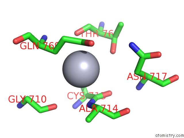
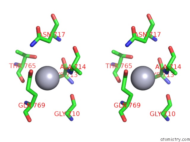
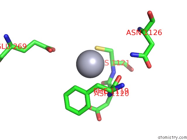
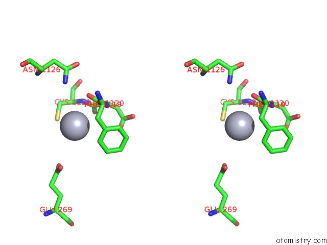
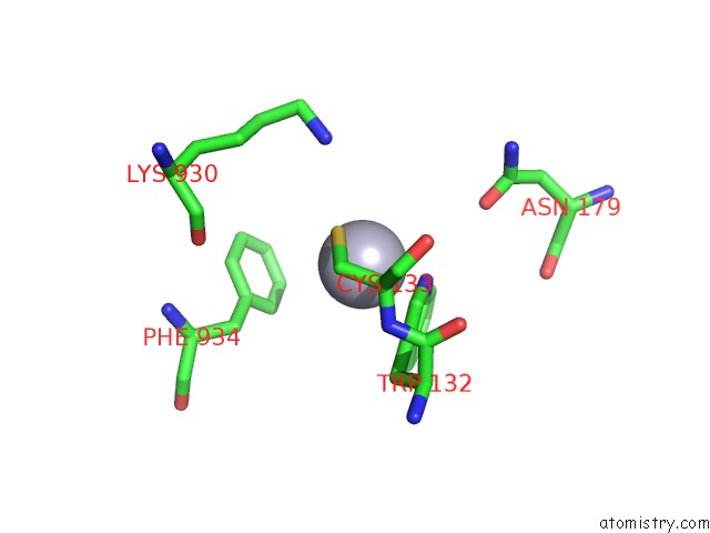
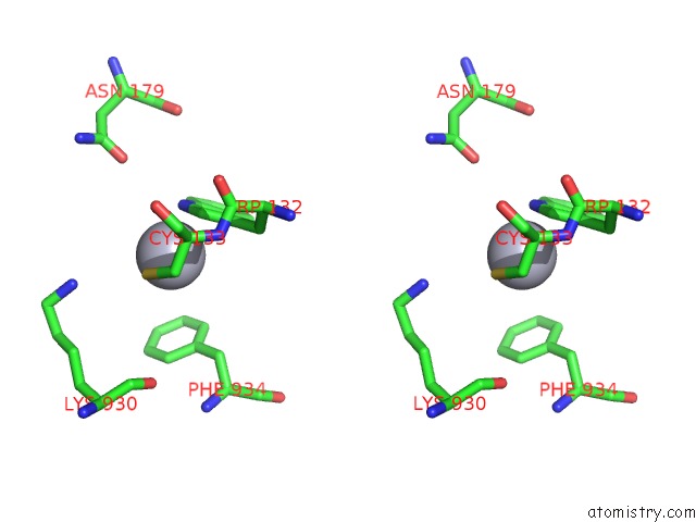
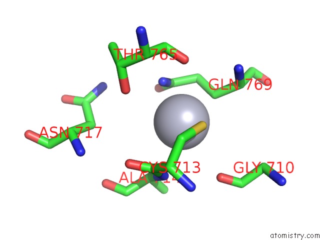
Mercury binding site 1 out of 12 in 3g5u
Go back to
Mercury binding site 1 out
of 12 in the Structure of P-Glycoprotein Reveals A Molecular Basis For Poly-Specific Drug Binding
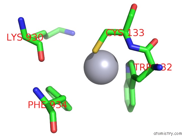
Mono view
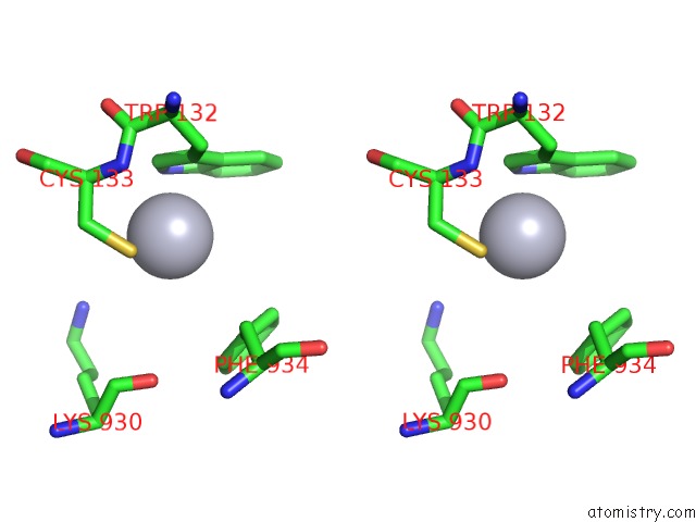
Stereo pair view
Mono view
Stereo pair view
A full contact list of Mercury with other atoms in the Hg binding
site number 1 of Structure of P-Glycoprotein Reveals A Molecular Basis For Poly-Specific Drug Binding within 5.0Å range:
|
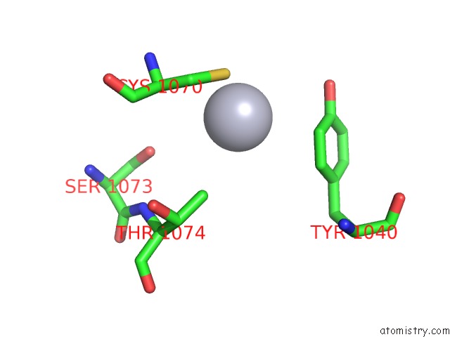
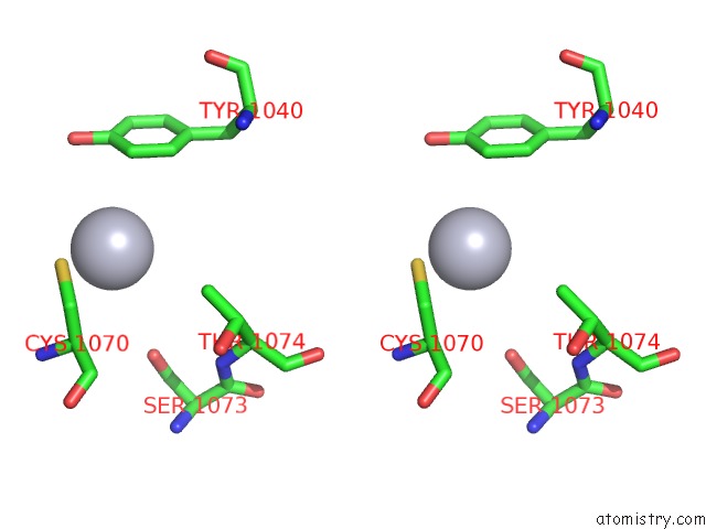
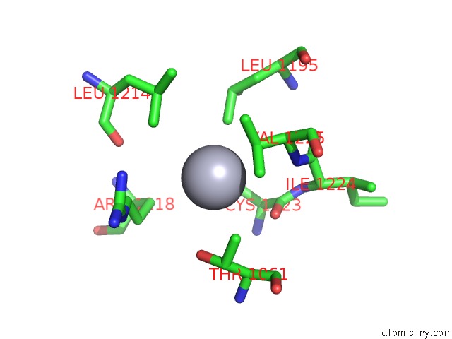
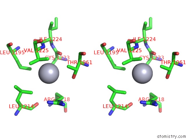
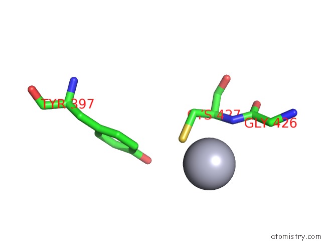
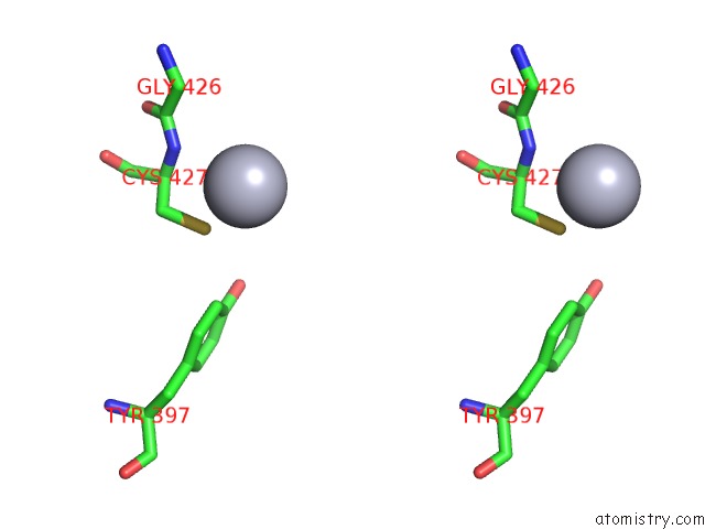
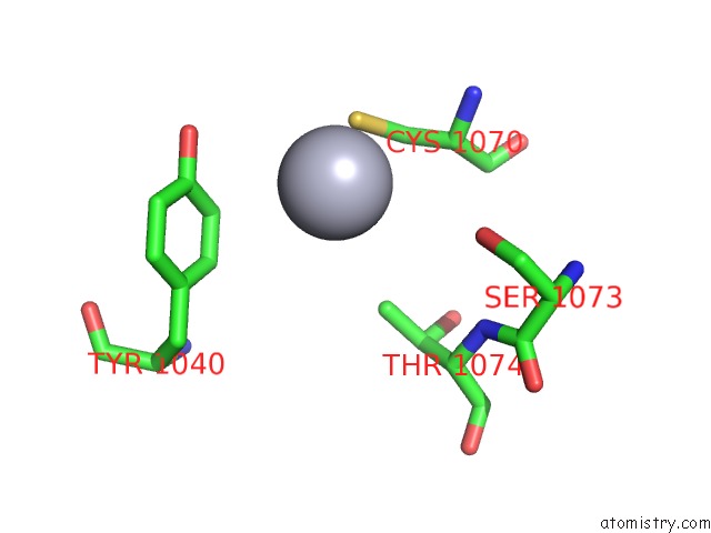
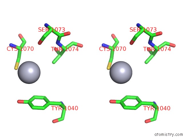
Mercury binding site 2 out of 12 in 3g5u
Go back to
Mercury binding site 2 out
of 12 in the Structure of P-Glycoprotein Reveals A Molecular Basis For Poly-Specific Drug Binding
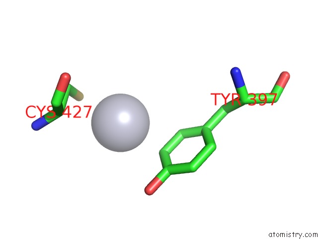
Mono view
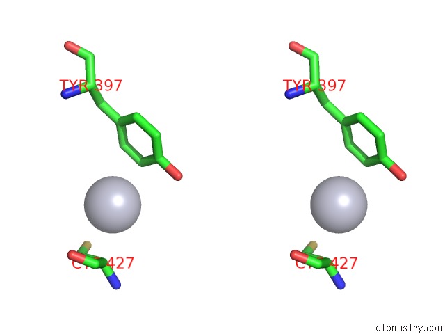
Stereo pair view
Mono view
Stereo pair view
A full contact list of Mercury with other atoms in the Hg binding
site number 2 of Structure of P-Glycoprotein Reveals A Molecular Basis For Poly-Specific Drug Binding within 5.0Å range:
|
Mercury binding site 3 out of 12 in 3g5u
Go back to
Mercury binding site 3 out
of 12 in the Structure of P-Glycoprotein Reveals A Molecular Basis For Poly-Specific Drug Binding
Mono view
Stereo pair view
Mono view
Stereo pair view
A full contact list of Mercury with other atoms in the Hg binding
site number 3 of Structure of P-Glycoprotein Reveals A Molecular Basis For Poly-Specific Drug Binding within 5.0Å range:
|
Mercury binding site 4 out of 12 in 3g5u
Go back to
Mercury binding site 4 out
of 12 in the Structure of P-Glycoprotein Reveals A Molecular Basis For Poly-Specific Drug Binding
Mono view
Stereo pair view
Mono view
Stereo pair view
A full contact list of Mercury with other atoms in the Hg binding
site number 4 of Structure of P-Glycoprotein Reveals A Molecular Basis For Poly-Specific Drug Binding within 5.0Å range:
|
Mercury binding site 5 out of 12 in 3g5u
Go back to
Mercury binding site 5 out
of 12 in the Structure of P-Glycoprotein Reveals A Molecular Basis For Poly-Specific Drug Binding
Mono view
Stereo pair view
Mono view
Stereo pair view
A full contact list of Mercury with other atoms in the Hg binding
site number 5 of Structure of P-Glycoprotein Reveals A Molecular Basis For Poly-Specific Drug Binding within 5.0Å range:
|
Mercury binding site 6 out of 12 in 3g5u
Go back to
Mercury binding site 6 out
of 12 in the Structure of P-Glycoprotein Reveals A Molecular Basis For Poly-Specific Drug Binding
Mono view
Stereo pair view
Mono view
Stereo pair view
A full contact list of Mercury with other atoms in the Hg binding
site number 6 of Structure of P-Glycoprotein Reveals A Molecular Basis For Poly-Specific Drug Binding within 5.0Å range:
|
Mercury binding site 7 out of 12 in 3g5u
Go back to
Mercury binding site 7 out
of 12 in the Structure of P-Glycoprotein Reveals A Molecular Basis For Poly-Specific Drug Binding
Mono view
Stereo pair view
Mono view
Stereo pair view
A full contact list of Mercury with other atoms in the Hg binding
site number 7 of Structure of P-Glycoprotein Reveals A Molecular Basis For Poly-Specific Drug Binding within 5.0Å range:
|
Mercury binding site 8 out of 12 in 3g5u
Go back to
Mercury binding site 8 out
of 12 in the Structure of P-Glycoprotein Reveals A Molecular Basis For Poly-Specific Drug Binding
Mono view
Stereo pair view
Mono view
Stereo pair view
A full contact list of Mercury with other atoms in the Hg binding
site number 8 of Structure of P-Glycoprotein Reveals A Molecular Basis For Poly-Specific Drug Binding within 5.0Å range:
|
Mercury binding site 9 out of 12 in 3g5u
Go back to
Mercury binding site 9 out
of 12 in the Structure of P-Glycoprotein Reveals A Molecular Basis For Poly-Specific Drug Binding
Mono view

Stereo pair view
Mono view
Stereo pair view
A full contact list of Mercury with other atoms in the Hg binding
site number 9 of Structure of P-Glycoprotein Reveals A Molecular Basis For Poly-Specific Drug Binding within 5.0Å range:
|
Mercury binding site 10 out of 12 in 3g5u
Go back to
Mercury binding site 10 out
of 12 in the Structure of P-Glycoprotein Reveals A Molecular Basis For Poly-Specific Drug Binding
Mono view
Stereo pair view
Mono view
Stereo pair view
A full contact list of Mercury with other atoms in the Hg binding
site number 10 of Structure of P-Glycoprotein Reveals A Molecular Basis For Poly-Specific Drug Binding within 5.0Å range:
|
Reference:
S.G.Aller,
J.Yu,
A.Ward,
Y.Weng,
S.Chittaboina,
R.Zhuo,
P.M.Harrell,
Y.T.Trinh,
Q.Zhang,
I.L.Urbatsch,
G.Chang.
Structure of P-Glycoprotein Reveals A Molecular Basis For Poly-Specific Drug Binding. Science V. 323 1718 2009.
ISSN: ISSN 0036-8075
PubMed: 19325113
DOI: 10.1126/SCIENCE.1168750
Page generated: Sun Aug 11 03:45:04 2024
ISSN: ISSN 0036-8075
PubMed: 19325113
DOI: 10.1126/SCIENCE.1168750
Last articles
Zn in 9MJ5Zn in 9HNW
Zn in 9G0L
Zn in 9FNE
Zn in 9DZN
Zn in 9E0I
Zn in 9D32
Zn in 9DAK
Zn in 8ZXC
Zn in 8ZUF