Mercury »
PDB 3bl0-3k7k »
3i34 »
Mercury in PDB 3i34: Proteinase K By Lb Nanotemplate Method After High X-Ray Dose on ID14-2 Beamline at Esrf
Enzymatic activity of Proteinase K By Lb Nanotemplate Method After High X-Ray Dose on ID14-2 Beamline at Esrf
All present enzymatic activity of Proteinase K By Lb Nanotemplate Method After High X-Ray Dose on ID14-2 Beamline at Esrf:
3.4.21.64;
3.4.21.64;
Protein crystallography data
The structure of Proteinase K By Lb Nanotemplate Method After High X-Ray Dose on ID14-2 Beamline at Esrf, PDB code: 3i34
was solved by
E.Pechkova,
S.K.Tripathi,
R.Ravelli,
S.Mcsweeney,
C.Nicolini,
with X-Ray Crystallography technique. A brief refinement statistics is given in the table below:
| Resolution Low / High (Å) | 23.99 / 1.00 |
| Space group | P 43 21 2 |
| Cell size a, b, c (Å), α, β, γ (°) | 67.865, 67.865, 102.335, 90.00, 90.00, 90.00 |
| R / Rfree (%) | 20.6 / 22 |
Other elements in 3i34:
The structure of Proteinase K By Lb Nanotemplate Method After High X-Ray Dose on ID14-2 Beamline at Esrf also contains other interesting chemical elements:
| Calcium | (Ca) | 1 atom |
Mercury Binding Sites:
The binding sites of Mercury atom in the Proteinase K By Lb Nanotemplate Method After High X-Ray Dose on ID14-2 Beamline at Esrf
(pdb code 3i34). This binding sites where shown within
5.0 Angstroms radius around Mercury atom.
In total 3 binding sites of Mercury where determined in the Proteinase K By Lb Nanotemplate Method After High X-Ray Dose on ID14-2 Beamline at Esrf, PDB code: 3i34:
Jump to Mercury binding site number: 1; 2; 3;
In total 3 binding sites of Mercury where determined in the Proteinase K By Lb Nanotemplate Method After High X-Ray Dose on ID14-2 Beamline at Esrf, PDB code: 3i34:
Jump to Mercury binding site number: 1; 2; 3;
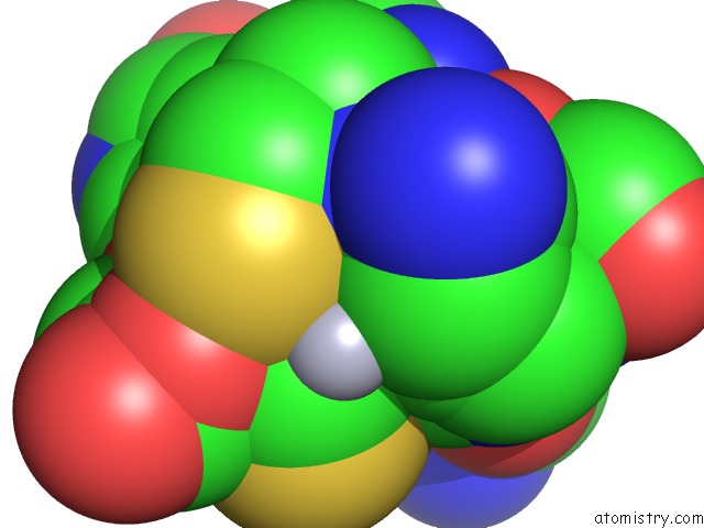
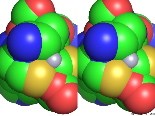
Mercury binding site 1 out of 3 in 3i34
Go back to
Mercury binding site 1 out
of 3 in the Proteinase K By Lb Nanotemplate Method After High X-Ray Dose on ID14-2 Beamline at Esrf
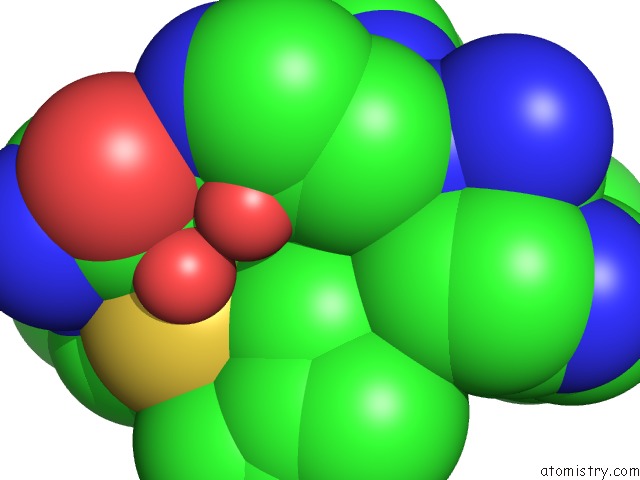
Mono view
Stereo pair view
Mono view
Stereo pair view
A full contact list of Mercury with other atoms in the Hg binding
site number 1 of Proteinase K By Lb Nanotemplate Method After High X-Ray Dose on ID14-2 Beamline at Esrf within 5.0Å range:
|
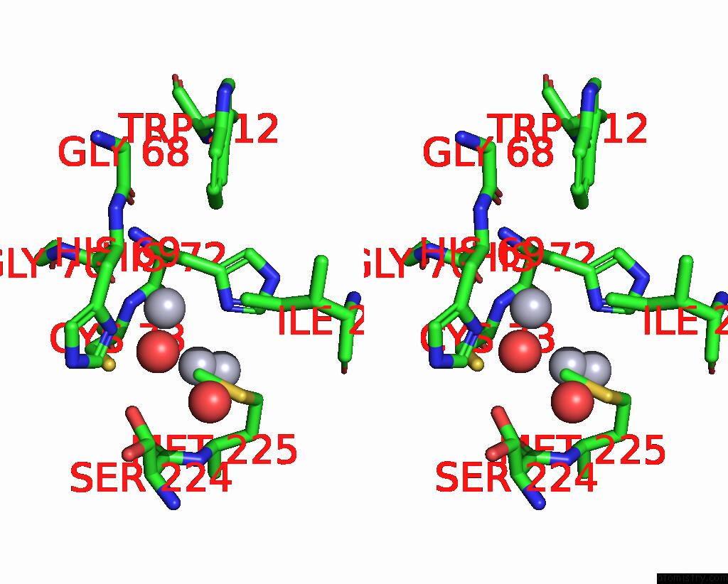
Mercury binding site 2 out of 3 in 3i34
Go back to
Mercury binding site 2 out
of 3 in the Proteinase K By Lb Nanotemplate Method After High X-Ray Dose on ID14-2 Beamline at Esrf
Mono view
Stereo pair view
Mono view
Stereo pair view
A full contact list of Mercury with other atoms in the Hg binding
site number 2 of Proteinase K By Lb Nanotemplate Method After High X-Ray Dose on ID14-2 Beamline at Esrf within 5.0Å range:
|
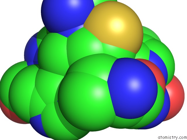
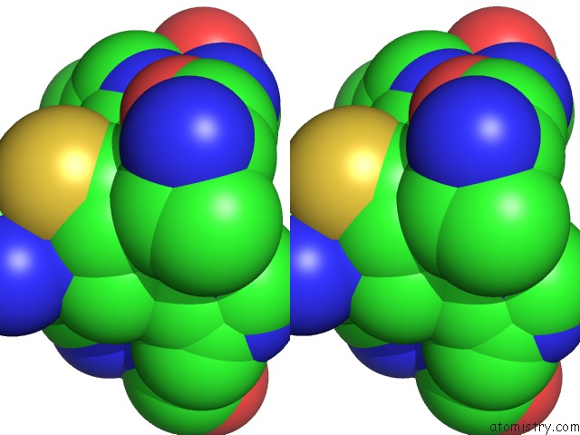
Mercury binding site 3 out of 3 in 3i34
Go back to
Mercury binding site 3 out
of 3 in the Proteinase K By Lb Nanotemplate Method After High X-Ray Dose on ID14-2 Beamline at Esrf
Mono view
Stereo pair view
Mono view
Stereo pair view
A full contact list of Mercury with other atoms in the Hg binding
site number 3 of Proteinase K By Lb Nanotemplate Method After High X-Ray Dose on ID14-2 Beamline at Esrf within 5.0Å range:
|
Reference:
E.Pechkova,
S.K.Tripathi,
R.Ravelli,
S.Mcsweeney,
C.Nicolini.
Radiation Damage Study of Proteinase K at ID14-2 Beamline at Esrf To Be Published.
Page generated: Fri Aug 8 10:09:51 2025
Last articles
I in 2OHUI in 2OHT
I in 2OHS
I in 2OHN
I in 2OHR
I in 2OHQ
I in 2OHP
I in 2OHK
I in 2OHL
I in 2O2B