Mercury »
PDB 3bl0-3k7k »
3igq »
Mercury in PDB 3igq: Crystal Structure of the Extracellular Domain of A Bacterial Pentameric Ligand-Gated Ion Channel
Protein crystallography data
The structure of Crystal Structure of the Extracellular Domain of A Bacterial Pentameric Ligand-Gated Ion Channel, PDB code: 3igq
was solved by
H.Nury,
M.Delarue,
with X-Ray Crystallography technique. A brief refinement statistics is given in the table below:
| Resolution Low / High (Å) | 25.00 / 2.30 |
| Space group | P 21 21 2 |
| Cell size a, b, c (Å), α, β, γ (°) | 84.450, 130.240, 113.590, 90.00, 90.00, 90.00 |
| R / Rfree (%) | 20.7 / 25.5 |
Other elements in 3igq:
The structure of Crystal Structure of the Extracellular Domain of A Bacterial Pentameric Ligand-Gated Ion Channel also contains other interesting chemical elements:
| Chlorine | (Cl) | 6 atoms |
| Sodium | (Na) | 6 atoms |
Mercury Binding Sites:
The binding sites of Mercury atom in the Crystal Structure of the Extracellular Domain of A Bacterial Pentameric Ligand-Gated Ion Channel
(pdb code 3igq). This binding sites where shown within
5.0 Angstroms radius around Mercury atom.
In total 6 binding sites of Mercury where determined in the Crystal Structure of the Extracellular Domain of A Bacterial Pentameric Ligand-Gated Ion Channel, PDB code: 3igq:
Jump to Mercury binding site number: 1; 2; 3; 4; 5; 6;
In total 6 binding sites of Mercury where determined in the Crystal Structure of the Extracellular Domain of A Bacterial Pentameric Ligand-Gated Ion Channel, PDB code: 3igq:
Jump to Mercury binding site number: 1; 2; 3; 4; 5; 6;
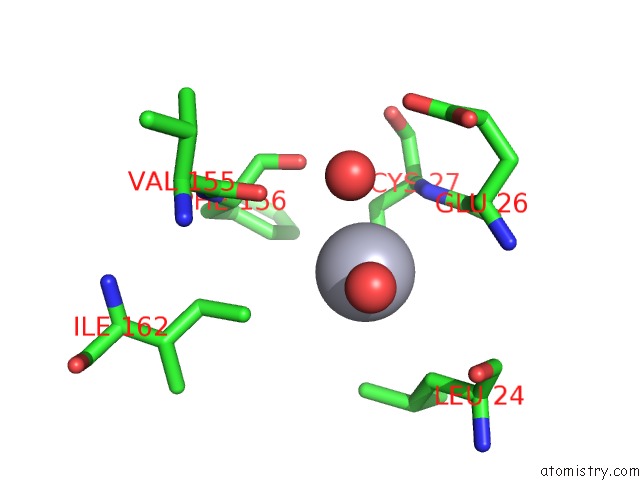
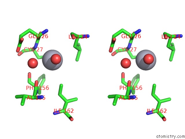
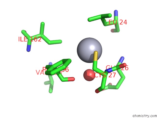
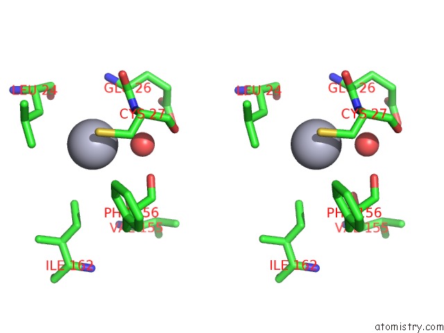
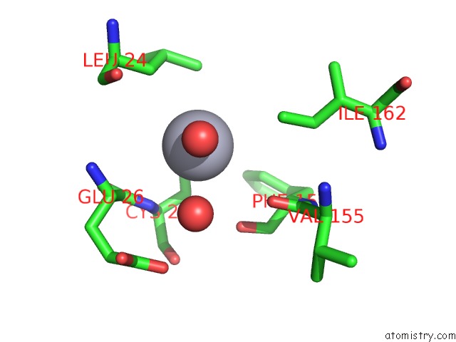
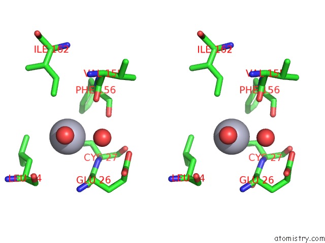
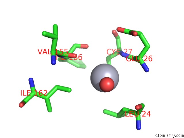
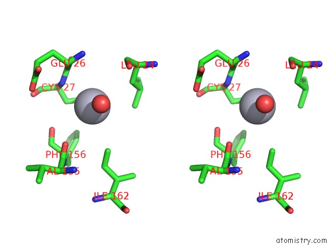
Mercury binding site 1 out of 6 in 3igq
Go back to
Mercury binding site 1 out
of 6 in the Crystal Structure of the Extracellular Domain of A Bacterial Pentameric Ligand-Gated Ion Channel
Mono view
Stereo pair view
Mono view
Stereo pair view
A full contact list of Mercury with other atoms in the Hg binding
site number 1 of Crystal Structure of the Extracellular Domain of A Bacterial Pentameric Ligand-Gated Ion Channel within 5.0Å range:
|
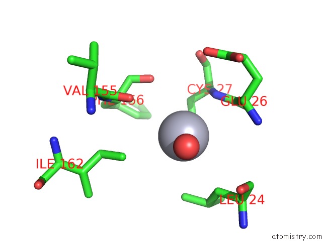
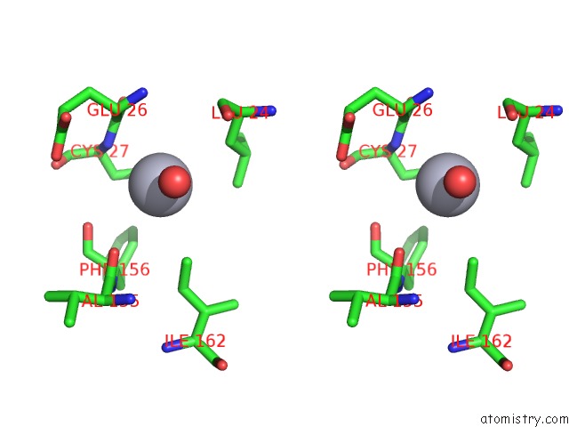
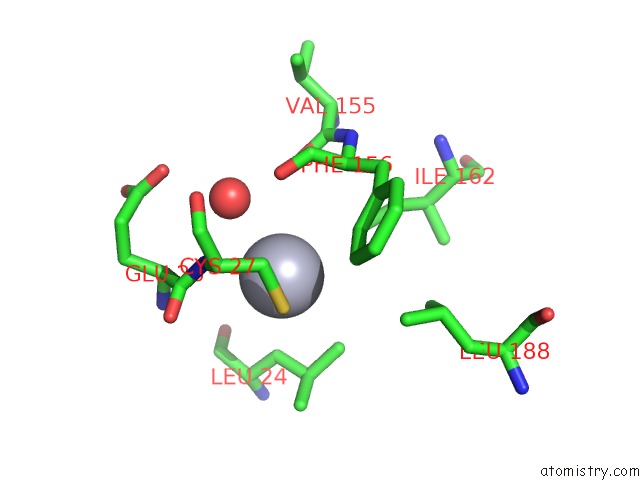
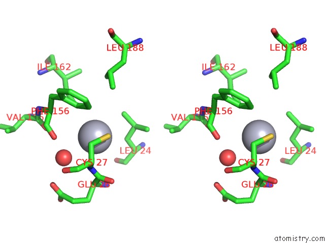
Mercury binding site 2 out of 6 in 3igq
Go back to
Mercury binding site 2 out
of 6 in the Crystal Structure of the Extracellular Domain of A Bacterial Pentameric Ligand-Gated Ion Channel
Mono view
Stereo pair view
Mono view
Stereo pair view
A full contact list of Mercury with other atoms in the Hg binding
site number 2 of Crystal Structure of the Extracellular Domain of A Bacterial Pentameric Ligand-Gated Ion Channel within 5.0Å range:
|
Mercury binding site 3 out of 6 in 3igq
Go back to
Mercury binding site 3 out
of 6 in the Crystal Structure of the Extracellular Domain of A Bacterial Pentameric Ligand-Gated Ion Channel
Mono view
Stereo pair view
Mono view
Stereo pair view
A full contact list of Mercury with other atoms in the Hg binding
site number 3 of Crystal Structure of the Extracellular Domain of A Bacterial Pentameric Ligand-Gated Ion Channel within 5.0Å range:
|
Mercury binding site 4 out of 6 in 3igq
Go back to
Mercury binding site 4 out
of 6 in the Crystal Structure of the Extracellular Domain of A Bacterial Pentameric Ligand-Gated Ion Channel
Mono view
Stereo pair view
Mono view
Stereo pair view
A full contact list of Mercury with other atoms in the Hg binding
site number 4 of Crystal Structure of the Extracellular Domain of A Bacterial Pentameric Ligand-Gated Ion Channel within 5.0Å range:
|
Mercury binding site 5 out of 6 in 3igq
Go back to
Mercury binding site 5 out
of 6 in the Crystal Structure of the Extracellular Domain of A Bacterial Pentameric Ligand-Gated Ion Channel
Mono view
Stereo pair view
Mono view
Stereo pair view
A full contact list of Mercury with other atoms in the Hg binding
site number 5 of Crystal Structure of the Extracellular Domain of A Bacterial Pentameric Ligand-Gated Ion Channel within 5.0Å range:
|
Mercury binding site 6 out of 6 in 3igq
Go back to
Mercury binding site 6 out
of 6 in the Crystal Structure of the Extracellular Domain of A Bacterial Pentameric Ligand-Gated Ion Channel
Mono view
Stereo pair view
Mono view
Stereo pair view
A full contact list of Mercury with other atoms in the Hg binding
site number 6 of Crystal Structure of the Extracellular Domain of A Bacterial Pentameric Ligand-Gated Ion Channel within 5.0Å range:
|
Reference:
H.Nury,
N.Bocquet,
C.Le Poupon,
B.Raynal,
A.Haouz,
P.-J.Corringer,
M.Delarue.
Crystal Structure of the Extracellular Domain of A Bacterial Ligand-Gated Ion Channel J.Mol.Biol. V. 395 1114 2010.
ISSN: ISSN 0022-2836
PubMed: 19917292
DOI: 10.1016/J.JMB.2009.11.024
Page generated: Sun Aug 11 03:50:07 2024
ISSN: ISSN 0022-2836
PubMed: 19917292
DOI: 10.1016/J.JMB.2009.11.024
Last articles
Cl in 3N9RCl in 3NB5
Cl in 3NA6
Cl in 3N9J
Cl in 3N9E
Cl in 3N9C
Cl in 3N9B
Cl in 3N8Y
Cl in 3N9A
Cl in 3N8U