Mercury »
PDB 3kbc-3wa8 »
3kbu »
Mercury in PDB 3kbu: Crystal Structure of the Ankyrin Binding Domain of Human Erythroid Beta Spectrin (Repeats 13-15) in Complex with the Spectrin Binding Domain of Human Erythroid Ankyrin (ZU5-Ank), Emts Derivative
Protein crystallography data
The structure of Crystal Structure of the Ankyrin Binding Domain of Human Erythroid Beta Spectrin (Repeats 13-15) in Complex with the Spectrin Binding Domain of Human Erythroid Ankyrin (ZU5-Ank), Emts Derivative, PDB code: 3kbu
was solved by
J.J.Ipsaro,
A.Mondragon,
with X-Ray Crystallography technique. A brief refinement statistics is given in the table below:
| Resolution Low / High (Å) | 37.82 / 2.75 |
| Space group | P 21 21 21 |
| Cell size a, b, c (Å), α, β, γ (°) | 90.130, 98.540, 137.930, 90.00, 90.00, 90.00 |
| R / Rfree (%) | 22.5 / 27.7 |
Mercury Binding Sites:
The binding sites of Mercury atom in the Crystal Structure of the Ankyrin Binding Domain of Human Erythroid Beta Spectrin (Repeats 13-15) in Complex with the Spectrin Binding Domain of Human Erythroid Ankyrin (ZU5-Ank), Emts Derivative
(pdb code 3kbu). This binding sites where shown within
5.0 Angstroms radius around Mercury atom.
In total 8 binding sites of Mercury where determined in the Crystal Structure of the Ankyrin Binding Domain of Human Erythroid Beta Spectrin (Repeats 13-15) in Complex with the Spectrin Binding Domain of Human Erythroid Ankyrin (ZU5-Ank), Emts Derivative, PDB code: 3kbu:
Jump to Mercury binding site number: 1; 2; 3; 4; 5; 6; 7; 8;
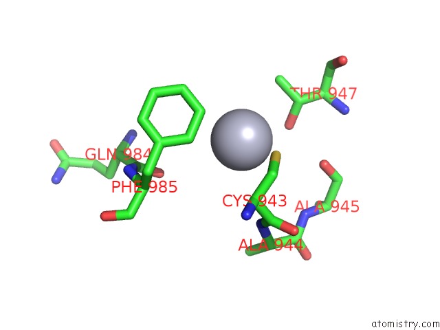
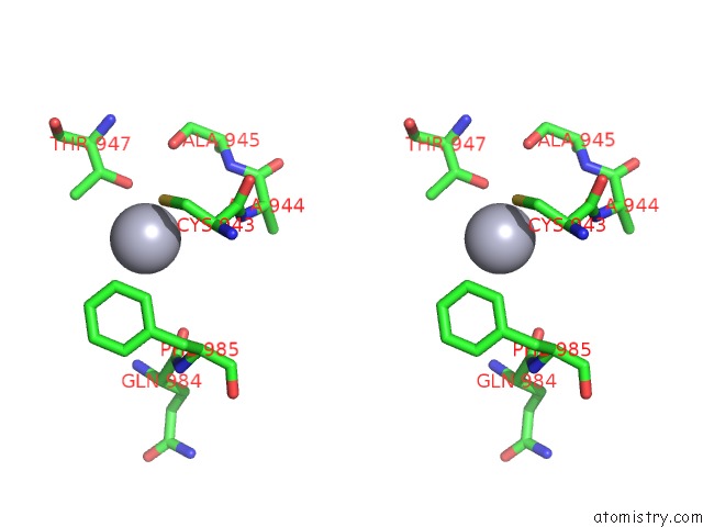
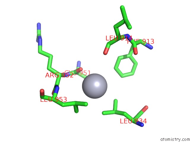
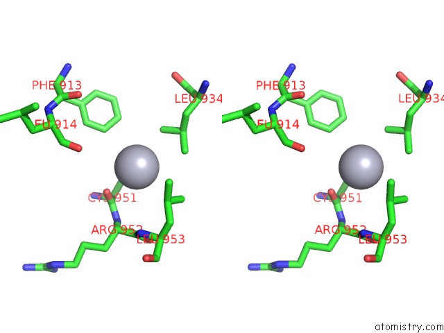
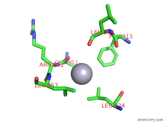
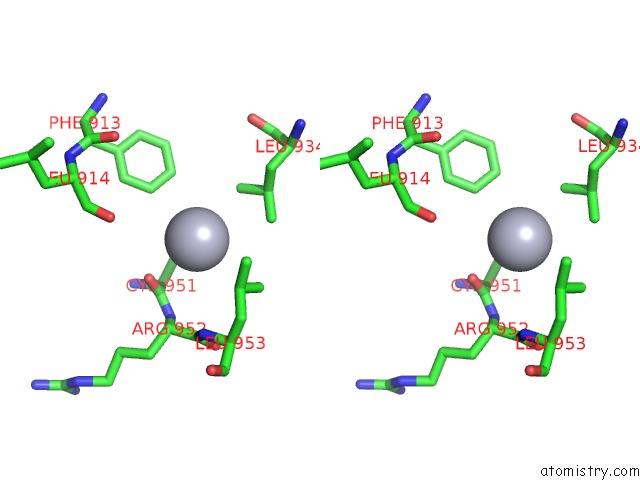
Mercury binding site 1 out of 8 in 3kbu
Go back to
Mercury binding site 1 out
of 8 in the Crystal Structure of the Ankyrin Binding Domain of Human Erythroid Beta Spectrin (Repeats 13-15) in Complex with the Spectrin Binding Domain of Human Erythroid Ankyrin (ZU5-Ank), Emts Derivative
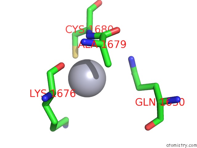
Mono view
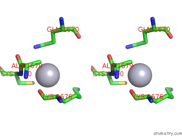
Stereo pair view
Mono view
Stereo pair view
A full contact list of Mercury with other atoms in the Hg binding
site number 1 of Crystal Structure of the Ankyrin Binding Domain of Human Erythroid Beta Spectrin (Repeats 13-15) in Complex with the Spectrin Binding Domain of Human Erythroid Ankyrin (ZU5-Ank), Emts Derivative within 5.0Å range:
|
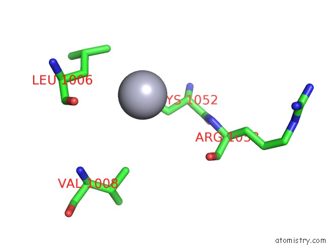
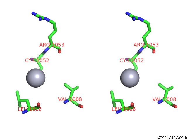
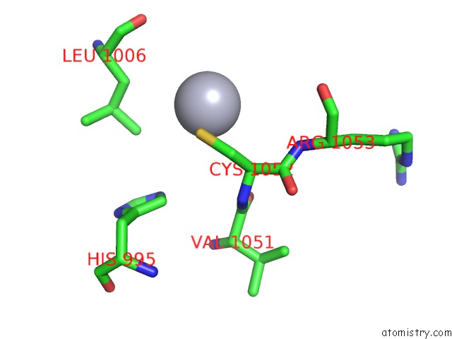
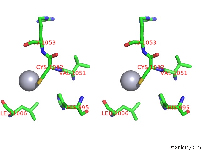
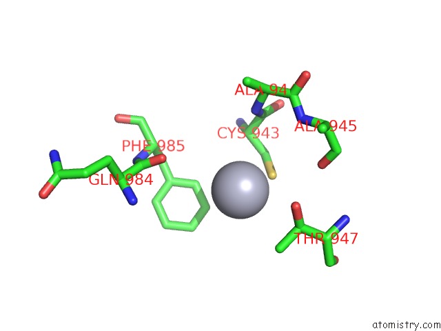
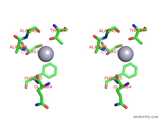
Mercury binding site 2 out of 8 in 3kbu
Go back to
Mercury binding site 2 out
of 8 in the Crystal Structure of the Ankyrin Binding Domain of Human Erythroid Beta Spectrin (Repeats 13-15) in Complex with the Spectrin Binding Domain of Human Erythroid Ankyrin (ZU5-Ank), Emts Derivative
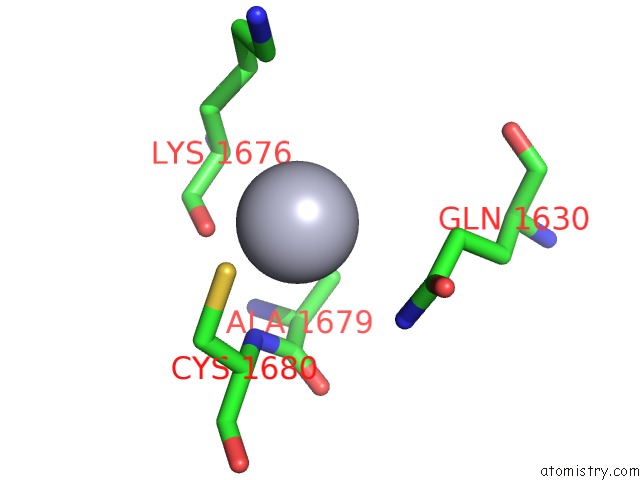
Mono view
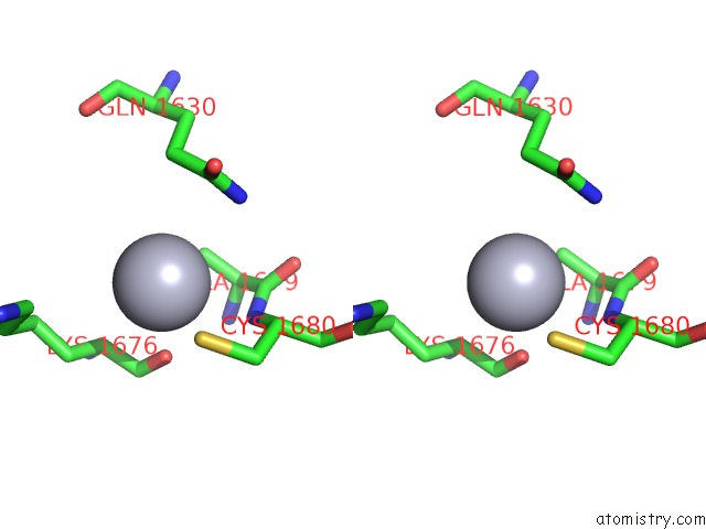
Stereo pair view
Mono view
Stereo pair view
A full contact list of Mercury with other atoms in the Hg binding
site number 2 of Crystal Structure of the Ankyrin Binding Domain of Human Erythroid Beta Spectrin (Repeats 13-15) in Complex with the Spectrin Binding Domain of Human Erythroid Ankyrin (ZU5-Ank), Emts Derivative within 5.0Å range:
|
Mercury binding site 3 out of 8 in 3kbu
Go back to
Mercury binding site 3 out
of 8 in the Crystal Structure of the Ankyrin Binding Domain of Human Erythroid Beta Spectrin (Repeats 13-15) in Complex with the Spectrin Binding Domain of Human Erythroid Ankyrin (ZU5-Ank), Emts Derivative
Mono view
Stereo pair view
Mono view
Stereo pair view
A full contact list of Mercury with other atoms in the Hg binding
site number 3 of Crystal Structure of the Ankyrin Binding Domain of Human Erythroid Beta Spectrin (Repeats 13-15) in Complex with the Spectrin Binding Domain of Human Erythroid Ankyrin (ZU5-Ank), Emts Derivative within 5.0Å range:
|
Mercury binding site 4 out of 8 in 3kbu
Go back to
Mercury binding site 4 out
of 8 in the Crystal Structure of the Ankyrin Binding Domain of Human Erythroid Beta Spectrin (Repeats 13-15) in Complex with the Spectrin Binding Domain of Human Erythroid Ankyrin (ZU5-Ank), Emts Derivative
Mono view
Stereo pair view
Mono view
Stereo pair view
A full contact list of Mercury with other atoms in the Hg binding
site number 4 of Crystal Structure of the Ankyrin Binding Domain of Human Erythroid Beta Spectrin (Repeats 13-15) in Complex with the Spectrin Binding Domain of Human Erythroid Ankyrin (ZU5-Ank), Emts Derivative within 5.0Å range:
|
Mercury binding site 5 out of 8 in 3kbu
Go back to
Mercury binding site 5 out
of 8 in the Crystal Structure of the Ankyrin Binding Domain of Human Erythroid Beta Spectrin (Repeats 13-15) in Complex with the Spectrin Binding Domain of Human Erythroid Ankyrin (ZU5-Ank), Emts Derivative
Mono view
Stereo pair view
Mono view
Stereo pair view
A full contact list of Mercury with other atoms in the Hg binding
site number 5 of Crystal Structure of the Ankyrin Binding Domain of Human Erythroid Beta Spectrin (Repeats 13-15) in Complex with the Spectrin Binding Domain of Human Erythroid Ankyrin (ZU5-Ank), Emts Derivative within 5.0Å range:
|
Mercury binding site 6 out of 8 in 3kbu
Go back to
Mercury binding site 6 out
of 8 in the Crystal Structure of the Ankyrin Binding Domain of Human Erythroid Beta Spectrin (Repeats 13-15) in Complex with the Spectrin Binding Domain of Human Erythroid Ankyrin (ZU5-Ank), Emts Derivative
Mono view
Stereo pair view
Mono view
Stereo pair view
A full contact list of Mercury with other atoms in the Hg binding
site number 6 of Crystal Structure of the Ankyrin Binding Domain of Human Erythroid Beta Spectrin (Repeats 13-15) in Complex with the Spectrin Binding Domain of Human Erythroid Ankyrin (ZU5-Ank), Emts Derivative within 5.0Å range:
|
Mercury binding site 7 out of 8 in 3kbu
Go back to
Mercury binding site 7 out
of 8 in the Crystal Structure of the Ankyrin Binding Domain of Human Erythroid Beta Spectrin (Repeats 13-15) in Complex with the Spectrin Binding Domain of Human Erythroid Ankyrin (ZU5-Ank), Emts Derivative
Mono view
Stereo pair view
Mono view
Stereo pair view
A full contact list of Mercury with other atoms in the Hg binding
site number 7 of Crystal Structure of the Ankyrin Binding Domain of Human Erythroid Beta Spectrin (Repeats 13-15) in Complex with the Spectrin Binding Domain of Human Erythroid Ankyrin (ZU5-Ank), Emts Derivative within 5.0Å range:
|
Mercury binding site 8 out of 8 in 3kbu
Go back to
Mercury binding site 8 out
of 8 in the Crystal Structure of the Ankyrin Binding Domain of Human Erythroid Beta Spectrin (Repeats 13-15) in Complex with the Spectrin Binding Domain of Human Erythroid Ankyrin (ZU5-Ank), Emts Derivative
Mono view
Stereo pair view
Mono view
Stereo pair view
A full contact list of Mercury with other atoms in the Hg binding
site number 8 of Crystal Structure of the Ankyrin Binding Domain of Human Erythroid Beta Spectrin (Repeats 13-15) in Complex with the Spectrin Binding Domain of Human Erythroid Ankyrin (ZU5-Ank), Emts Derivative within 5.0Å range:
|
Reference:
J.J.Ipsaro,
A.Mondragon.
Structural Basis For Spectrin Recognition By Ankyrin. Blood V. 115 4093 2010.
ISSN: ISSN 0006-4971
PubMed: 20101027
DOI: 10.1182/BLOOD-2009-11-255604
Page generated: Sun Aug 11 04:00:31 2024
ISSN: ISSN 0006-4971
PubMed: 20101027
DOI: 10.1182/BLOOD-2009-11-255604
Last articles
F in 7L4KF in 7L4F
F in 7L26
F in 7L4H
F in 7L4C
F in 7L24
F in 7L25
F in 7L1H
F in 7L0Z
F in 7L07