Mercury »
PDB 3kbc-3wa8 »
3ldi »
Mercury in PDB 3ldi: Crystal Structure of Aprotinin in Complex with Sucrose Octasulfate: Unusual Interactions and Implication For Heparin Binding
Protein crystallography data
The structure of Crystal Structure of Aprotinin in Complex with Sucrose Octasulfate: Unusual Interactions and Implication For Heparin Binding, PDB code: 3ldi
was solved by
I.S.Yang,
T.G.Kim,
B.S.Park,
K.H.Kim,
with X-Ray Crystallography technique. A brief refinement statistics is given in the table below:
| Resolution Low / High (Å) | 34.77 / 2.20 |
| Space group | P 63 2 2 |
| Cell size a, b, c (Å), α, β, γ (°) | 119.080, 119.080, 110.794, 90.00, 90.00, 120.00 |
| R / Rfree (%) | 20.6 / 23.4 |
Mercury Binding Sites:
The binding sites of Mercury atom in the Crystal Structure of Aprotinin in Complex with Sucrose Octasulfate: Unusual Interactions and Implication For Heparin Binding
(pdb code 3ldi). This binding sites where shown within
5.0 Angstroms radius around Mercury atom.
In total 5 binding sites of Mercury where determined in the Crystal Structure of Aprotinin in Complex with Sucrose Octasulfate: Unusual Interactions and Implication For Heparin Binding, PDB code: 3ldi:
Jump to Mercury binding site number: 1; 2; 3; 4; 5;
In total 5 binding sites of Mercury where determined in the Crystal Structure of Aprotinin in Complex with Sucrose Octasulfate: Unusual Interactions and Implication For Heparin Binding, PDB code: 3ldi:
Jump to Mercury binding site number: 1; 2; 3; 4; 5;
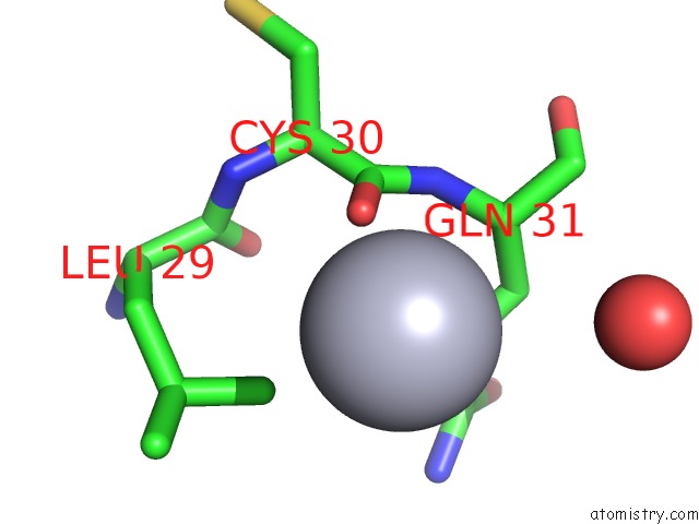
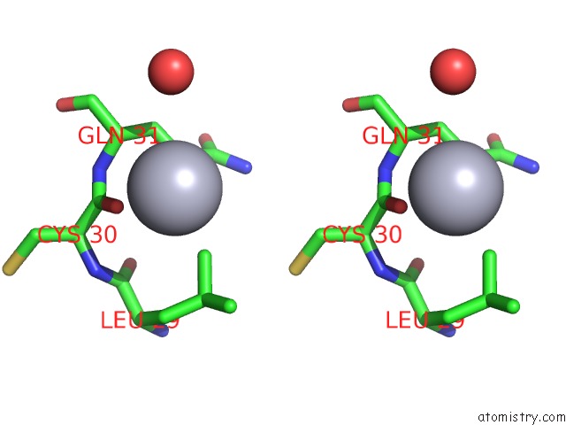
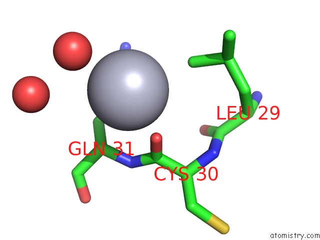
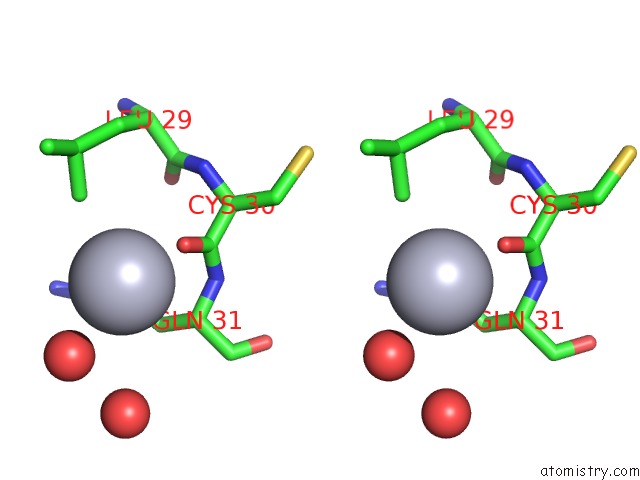
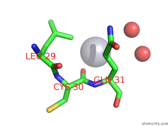
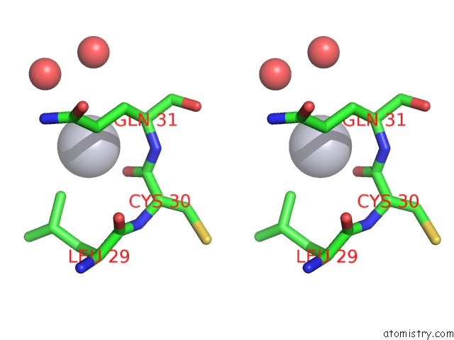
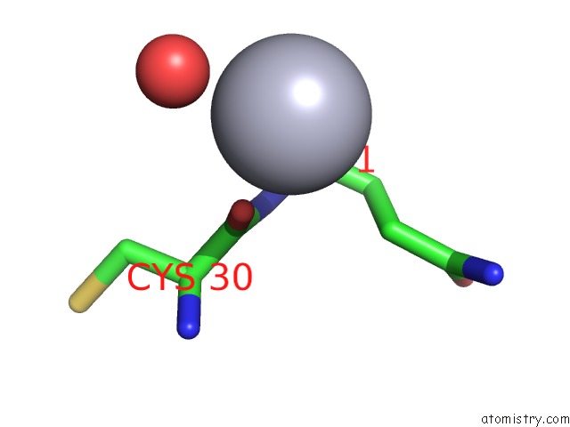
Mercury binding site 1 out of 5 in 3ldi
Go back to
Mercury binding site 1 out
of 5 in the Crystal Structure of Aprotinin in Complex with Sucrose Octasulfate: Unusual Interactions and Implication For Heparin Binding
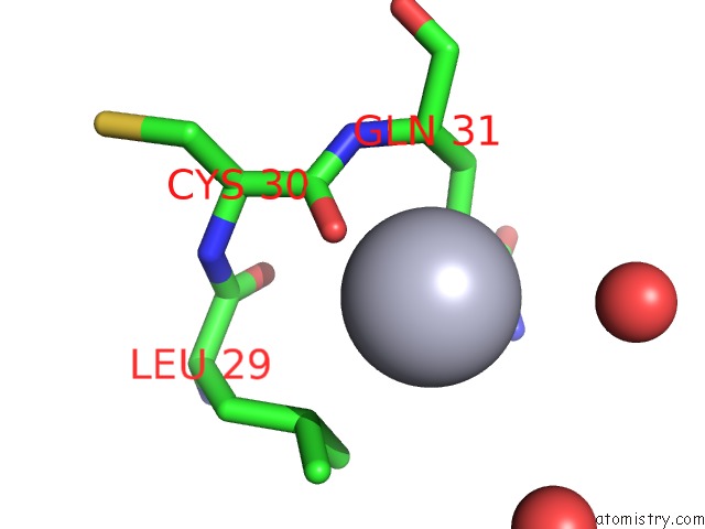
Mono view
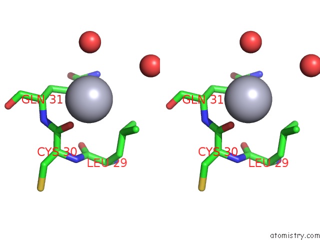
Stereo pair view
Mono view
Stereo pair view
A full contact list of Mercury with other atoms in the Hg binding
site number 1 of Crystal Structure of Aprotinin in Complex with Sucrose Octasulfate: Unusual Interactions and Implication For Heparin Binding within 5.0Å range:
|
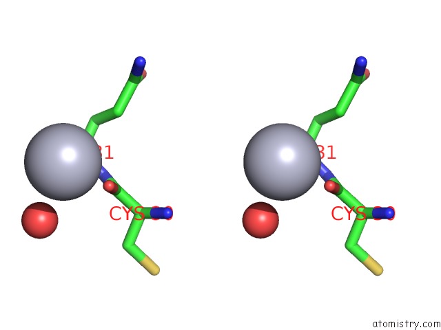
Mercury binding site 2 out of 5 in 3ldi
Go back to
Mercury binding site 2 out
of 5 in the Crystal Structure of Aprotinin in Complex with Sucrose Octasulfate: Unusual Interactions and Implication For Heparin Binding
Mono view
Stereo pair view
Mono view
Stereo pair view
A full contact list of Mercury with other atoms in the Hg binding
site number 2 of Crystal Structure of Aprotinin in Complex with Sucrose Octasulfate: Unusual Interactions and Implication For Heparin Binding within 5.0Å range:
|
Mercury binding site 3 out of 5 in 3ldi
Go back to
Mercury binding site 3 out
of 5 in the Crystal Structure of Aprotinin in Complex with Sucrose Octasulfate: Unusual Interactions and Implication For Heparin Binding
Mono view
Stereo pair view
Mono view
Stereo pair view
A full contact list of Mercury with other atoms in the Hg binding
site number 3 of Crystal Structure of Aprotinin in Complex with Sucrose Octasulfate: Unusual Interactions and Implication For Heparin Binding within 5.0Å range:
|
Mercury binding site 4 out of 5 in 3ldi
Go back to
Mercury binding site 4 out
of 5 in the Crystal Structure of Aprotinin in Complex with Sucrose Octasulfate: Unusual Interactions and Implication For Heparin Binding
Mono view
Stereo pair view
Mono view
Stereo pair view
A full contact list of Mercury with other atoms in the Hg binding
site number 4 of Crystal Structure of Aprotinin in Complex with Sucrose Octasulfate: Unusual Interactions and Implication For Heparin Binding within 5.0Å range:
|
Mercury binding site 5 out of 5 in 3ldi
Go back to
Mercury binding site 5 out
of 5 in the Crystal Structure of Aprotinin in Complex with Sucrose Octasulfate: Unusual Interactions and Implication For Heparin Binding
Mono view
Stereo pair view
Mono view
Stereo pair view
A full contact list of Mercury with other atoms in the Hg binding
site number 5 of Crystal Structure of Aprotinin in Complex with Sucrose Octasulfate: Unusual Interactions and Implication For Heparin Binding within 5.0Å range:
|
Reference:
I.S.Yang,
T.G.Kim,
B.S.Park,
K.J.Cho,
J.-H.Lee,
Y.Park,
K.H.Kim.
Crystal Structures of Aprotinin and Its Complex with Sucrose Octasulfate Reveal Multiple Modes of Interactions with Implications For Heparin Binding Biochem.Biophys.Res.Commun. V. 397 429 2010.
ISSN: ISSN 0006-291X
PubMed: 20529698
DOI: 10.1016/J.BBRC.2010.05.113
Page generated: Fri Aug 8 10:13:24 2025
ISSN: ISSN 0006-291X
PubMed: 20529698
DOI: 10.1016/J.BBRC.2010.05.113
Last articles
I in 2OF0I in 2JJS
I in 2J7P
I in 2NOO
I in 2HRA
I in 2NLL
I in 2NDO
I in 2L65
I in 2IZQ
I in 2IWK