Mercury »
PDB 3kbc-3wa8 »
3myz »
Mercury in PDB 3myz: Protein Induced Photophysical Changes to the Amyloid Indicator Dye, Thioflavin T
Protein crystallography data
The structure of Protein Induced Photophysical Changes to the Amyloid Indicator Dye, Thioflavin T, PDB code: 3myz
was solved by
L.S.Wolfe,
M.F.Calabrese,
A.Nath,
D.V.Blaho,
A.D.Miranker,
Y.Xiong,
with X-Ray Crystallography technique. A brief refinement statistics is given in the table below:
| Resolution Low / High (Å) | 29.27 / 1.60 |
| Space group | P 1 21 1 |
| Cell size a, b, c (Å), α, β, γ (°) | 48.349, 28.467, 74.983, 90.00, 91.42, 90.00 |
| R / Rfree (%) | 18.9 / 22.3 |
Mercury Binding Sites:
The binding sites of Mercury atom in the Protein Induced Photophysical Changes to the Amyloid Indicator Dye, Thioflavin T
(pdb code 3myz). This binding sites where shown within
5.0 Angstroms radius around Mercury atom.
In total only one binding site of Mercury was determined in the Protein Induced Photophysical Changes to the Amyloid Indicator Dye, Thioflavin T, PDB code: 3myz:
In total only one binding site of Mercury was determined in the Protein Induced Photophysical Changes to the Amyloid Indicator Dye, Thioflavin T, PDB code: 3myz:
Mercury binding site 1 out of 1 in 3myz
Go back to
Mercury binding site 1 out
of 1 in the Protein Induced Photophysical Changes to the Amyloid Indicator Dye, Thioflavin T
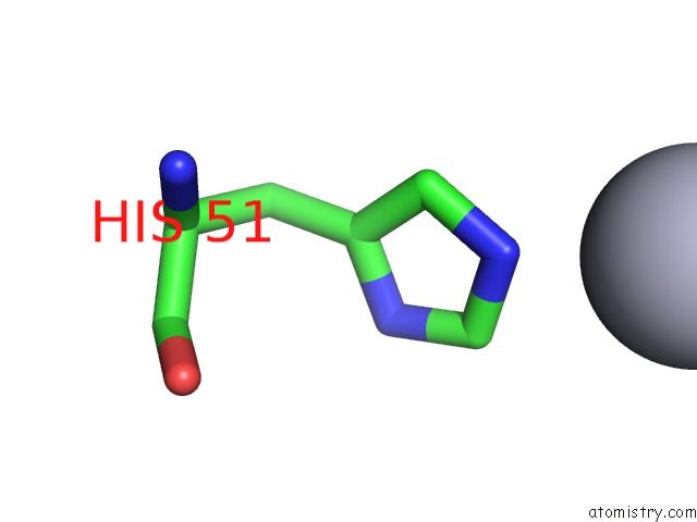
Mono view
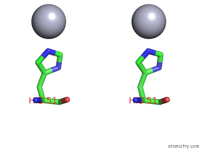
Stereo pair view
Mono view
Stereo pair view
A full contact list of Mercury with other atoms in the Hg binding
site number 1 of Protein Induced Photophysical Changes to the Amyloid Indicator Dye, Thioflavin T within 5.0Å range:
|
Reference:
L.S.Wolfe,
M.F.Calabrese,
A.Nath,
D.V.Blaho,
A.D.Miranker,
Y.Xiong.
Protein-Induced Photophysical Changes to the Amyloid Indicator Dye Thioflavin T. Proc.Natl.Acad.Sci.Usa V. 107 16863 2010.
ISSN: ISSN 0027-8424
PubMed: 20826442
DOI: 10.1073/PNAS.1002867107
Page generated: Fri Aug 8 10:14:24 2025
ISSN: ISSN 0027-8424
PubMed: 20826442
DOI: 10.1073/PNAS.1002867107
Last articles
I in 4PVGI in 4PGC
I in 4PNS
I in 4P4Z
I in 4P1E
I in 4P4X
I in 4P4Y
I in 4P4W
I in 4OA5
I in 4OWA