Mercury »
PDB 3kbc-3wa8 »
3oax »
Mercury in PDB 3oax: Crystal Structure of Bovine Rhodopsin with Beta-Ionone
Protein crystallography data
The structure of Crystal Structure of Bovine Rhodopsin with Beta-Ionone, PDB code: 3oax
was solved by
C.L.Makino,
C.K.Riley,
J.Looney,
R.K.Crouch,
T.Okada,
with X-Ray Crystallography technique. A brief refinement statistics is given in the table below:
| Resolution Low / High (Å) | 50.00 / 2.60 |
| Space group | P 41 |
| Cell size a, b, c (Å), α, β, γ (°) | 96.990, 96.990, 149.800, 90.00, 90.00, 90.00 |
| R / Rfree (%) | 22.4 / 26.2 |
Other elements in 3oax:
The structure of Crystal Structure of Bovine Rhodopsin with Beta-Ionone also contains other interesting chemical elements:
| Zinc | (Zn) | 7 atoms |
Mercury Binding Sites:
The binding sites of Mercury atom in the Crystal Structure of Bovine Rhodopsin with Beta-Ionone
(pdb code 3oax). This binding sites where shown within
5.0 Angstroms radius around Mercury atom.
In total 6 binding sites of Mercury where determined in the Crystal Structure of Bovine Rhodopsin with Beta-Ionone, PDB code: 3oax:
Jump to Mercury binding site number: 1; 2; 3; 4; 5; 6;
In total 6 binding sites of Mercury where determined in the Crystal Structure of Bovine Rhodopsin with Beta-Ionone, PDB code: 3oax:
Jump to Mercury binding site number: 1; 2; 3; 4; 5; 6;
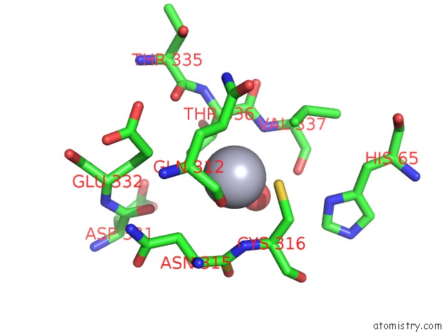
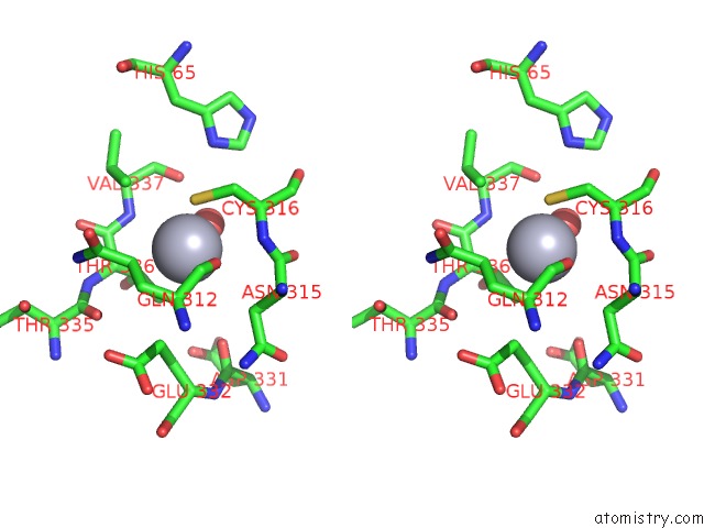
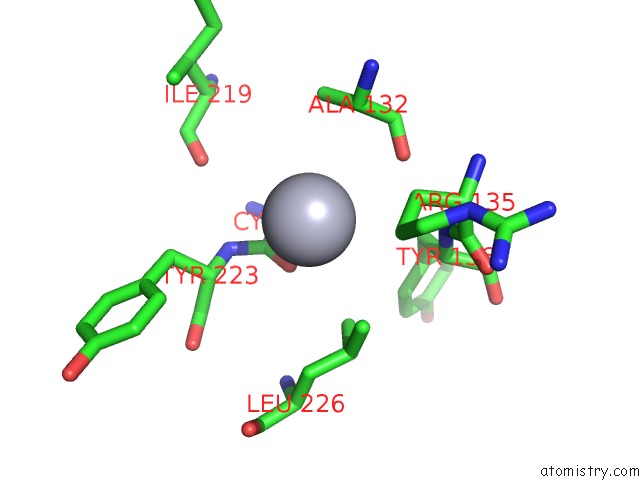
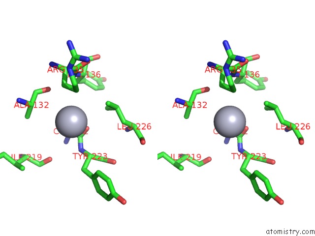
Mercury binding site 1 out of 6 in 3oax
Go back to
Mercury binding site 1 out
of 6 in the Crystal Structure of Bovine Rhodopsin with Beta-Ionone
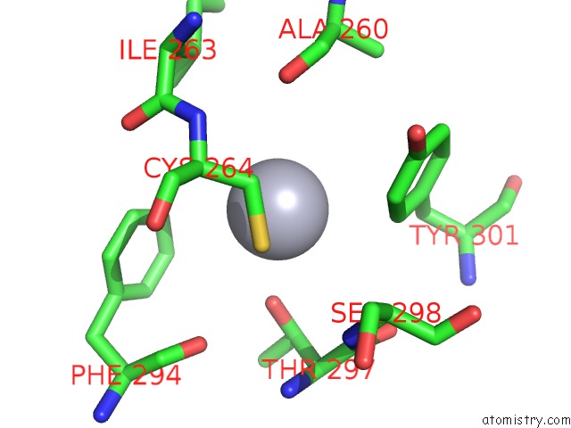
Mono view
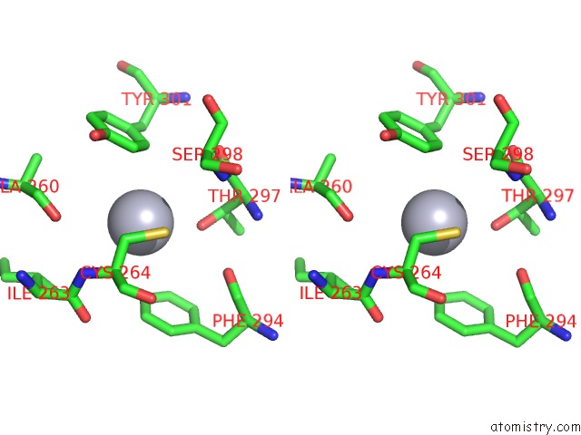
Stereo pair view
Mono view
Stereo pair view
A full contact list of Mercury with other atoms in the Hg binding
site number 1 of Crystal Structure of Bovine Rhodopsin with Beta-Ionone within 5.0Å range:
|
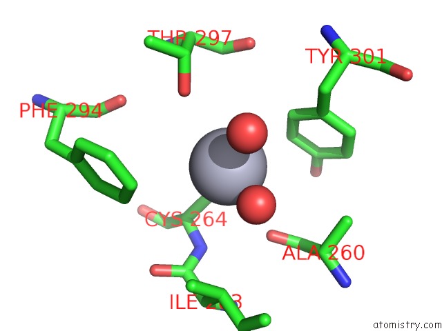
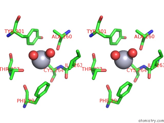
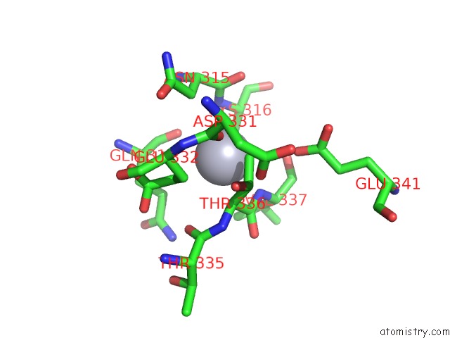
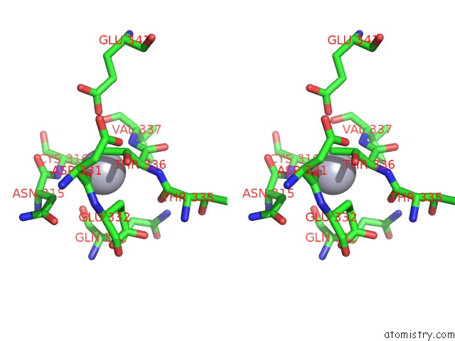
Mercury binding site 2 out of 6 in 3oax
Go back to
Mercury binding site 2 out
of 6 in the Crystal Structure of Bovine Rhodopsin with Beta-Ionone
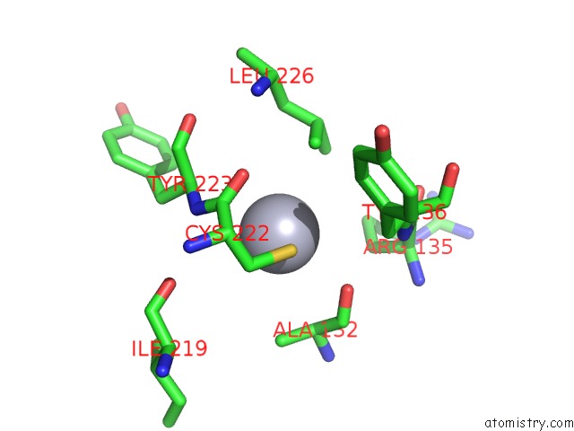
Mono view
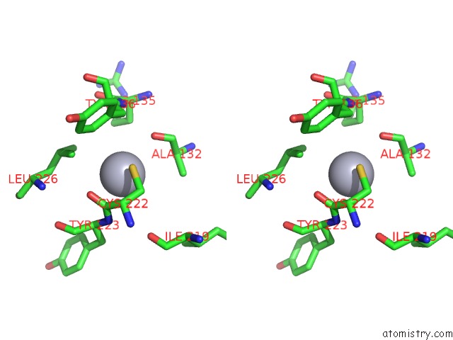
Stereo pair view
Mono view
Stereo pair view
A full contact list of Mercury with other atoms in the Hg binding
site number 2 of Crystal Structure of Bovine Rhodopsin with Beta-Ionone within 5.0Å range:
|
Mercury binding site 3 out of 6 in 3oax
Go back to
Mercury binding site 3 out
of 6 in the Crystal Structure of Bovine Rhodopsin with Beta-Ionone
Mono view
Stereo pair view
Mono view
Stereo pair view
A full contact list of Mercury with other atoms in the Hg binding
site number 3 of Crystal Structure of Bovine Rhodopsin with Beta-Ionone within 5.0Å range:
|
Mercury binding site 4 out of 6 in 3oax
Go back to
Mercury binding site 4 out
of 6 in the Crystal Structure of Bovine Rhodopsin with Beta-Ionone
Mono view
Stereo pair view
Mono view
Stereo pair view
A full contact list of Mercury with other atoms in the Hg binding
site number 4 of Crystal Structure of Bovine Rhodopsin with Beta-Ionone within 5.0Å range:
|
Mercury binding site 5 out of 6 in 3oax
Go back to
Mercury binding site 5 out
of 6 in the Crystal Structure of Bovine Rhodopsin with Beta-Ionone
Mono view
Stereo pair view
Mono view
Stereo pair view
A full contact list of Mercury with other atoms in the Hg binding
site number 5 of Crystal Structure of Bovine Rhodopsin with Beta-Ionone within 5.0Å range:
|
Mercury binding site 6 out of 6 in 3oax
Go back to
Mercury binding site 6 out
of 6 in the Crystal Structure of Bovine Rhodopsin with Beta-Ionone
Mono view
Stereo pair view
Mono view
Stereo pair view
A full contact list of Mercury with other atoms in the Hg binding
site number 6 of Crystal Structure of Bovine Rhodopsin with Beta-Ionone within 5.0Å range:
|
Reference:
C.L.Makino,
C.K.Riley,
J.Looney,
R.K.Crouch,
T.Okada.
Binding of More Than One Retinoid to Visual Opsins Biophys.J. V. 99 2366 2010.
ISSN: ISSN 0006-3495
PubMed: 20923672
DOI: 10.1016/J.BPJ.2010.08.003
Page generated: Sun Aug 11 04:06:07 2024
ISSN: ISSN 0006-3495
PubMed: 20923672
DOI: 10.1016/J.BPJ.2010.08.003
Last articles
Cl in 3NXSCl in 3NWB
Cl in 3NW9
Cl in 3NWN
Cl in 3NWE
Cl in 3NWD
Cl in 3NVS
Cl in 3NW8
Cl in 3NVR
Cl in 3NVU