Mercury »
PDB 3kbc-3wa8 »
3rx6 »
Mercury in PDB 3rx6: Crystal Structure of Polarity Suppression Protein From Enterobacteria Phage P4
Protein crystallography data
The structure of Crystal Structure of Polarity Suppression Protein From Enterobacteria Phage P4, PDB code: 3rx6
was solved by
R.Banerjee,
S.Nath,
S.Khamrui,
R.Sen,
U.Sen,
with X-Ray Crystallography technique. A brief refinement statistics is given in the table below:
| Resolution Low / High (Å) | 47.04 / 2.04 |
| Space group | I 4 2 2 |
| Cell size a, b, c (Å), α, β, γ (°) | 148.750, 148.750, 63.370, 90.00, 90.00, 90.00 |
| R / Rfree (%) | 19.2 / 21.9 |
Other elements in 3rx6:
The structure of Crystal Structure of Polarity Suppression Protein From Enterobacteria Phage P4 also contains other interesting chemical elements:
| Iodine | (I) | 1 atom |
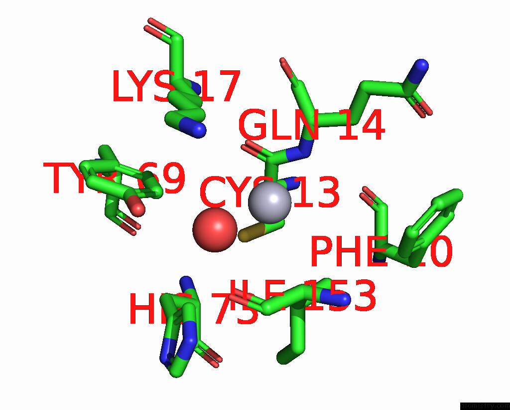
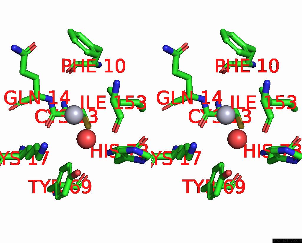
Mercury Binding Sites:
The binding sites of Mercury atom in the Crystal Structure of Polarity Suppression Protein From Enterobacteria Phage P4
(pdb code 3rx6). This binding sites where shown within
5.0 Angstroms radius around Mercury atom.
In total only one binding site of Mercury was determined in the Crystal Structure of Polarity Suppression Protein From Enterobacteria Phage P4, PDB code: 3rx6:
In total only one binding site of Mercury was determined in the Crystal Structure of Polarity Suppression Protein From Enterobacteria Phage P4, PDB code: 3rx6:
Mercury binding site 1 out of 1 in 3rx6
Go back to
Mercury binding site 1 out
of 1 in the Crystal Structure of Polarity Suppression Protein From Enterobacteria Phage P4
Mono view
Stereo pair view
Mono view
Stereo pair view
A full contact list of Mercury with other atoms in the Hg binding
site number 1 of Crystal Structure of Polarity Suppression Protein From Enterobacteria Phage P4 within 5.0Å range:
|
Reference:
R.Banerjee,
S.Nath,
A.Ranjan,
S.Khamrui,
B.Pani,
R.Sen,
U.Sen.
The First Structure of Polarity Suppression Protein, Psu From Enterobacteria Phage P4, Reveals A Novel Fold and A Knotted Dimer J.Biol.Chem. V. 287 44667 2012.
ISSN: ISSN 0021-9258
PubMed: 23150672
DOI: 10.1074/JBC.M112.423202
Page generated: Sun Aug 11 04:08:54 2024
ISSN: ISSN 0021-9258
PubMed: 23150672
DOI: 10.1074/JBC.M112.423202
Last articles
Zn in 9J0NZn in 9J0O
Zn in 9J0P
Zn in 9FJX
Zn in 9EKB
Zn in 9C0F
Zn in 9CAH
Zn in 9CH0
Zn in 9CH3
Zn in 9CH1