Mercury »
PDB 3wee-4ia4 »
4fvo »
Mercury in PDB 4fvo: Carbonic Anhydrase II in Complex with N-[(2E)-3,4-Dihydroquinazolin- 2(1H)-Ylidene]Sulfuric Diamide
Enzymatic activity of Carbonic Anhydrase II in Complex with N-[(2E)-3,4-Dihydroquinazolin- 2(1H)-Ylidene]Sulfuric Diamide
All present enzymatic activity of Carbonic Anhydrase II in Complex with N-[(2E)-3,4-Dihydroquinazolin- 2(1H)-Ylidene]Sulfuric Diamide:
4.2.1.1;
4.2.1.1;
Protein crystallography data
The structure of Carbonic Anhydrase II in Complex with N-[(2E)-3,4-Dihydroquinazolin- 2(1H)-Ylidene]Sulfuric Diamide, PDB code: 4fvo
was solved by
A.Di Pizio,
A.Heine,
G.Klebe,
with X-Ray Crystallography technique. A brief refinement statistics is given in the table below:
| Resolution Low / High (Å) | 30.00 / 1.05 |
| Space group | P 1 21 1 |
| Cell size a, b, c (Å), α, β, γ (°) | 42.139, 41.457, 72.169, 90.00, 104.21, 90.00 |
| R / Rfree (%) | 14 / 17 |
Other elements in 4fvo:
The structure of Carbonic Anhydrase II in Complex with N-[(2E)-3,4-Dihydroquinazolin- 2(1H)-Ylidene]Sulfuric Diamide also contains other interesting chemical elements:
| Zinc | (Zn) | 1 atom |
Mercury Binding Sites:
The binding sites of Mercury atom in the Carbonic Anhydrase II in Complex with N-[(2E)-3,4-Dihydroquinazolin- 2(1H)-Ylidene]Sulfuric Diamide
(pdb code 4fvo). This binding sites where shown within
5.0 Angstroms radius around Mercury atom.
In total only one binding site of Mercury was determined in the Carbonic Anhydrase II in Complex with N-[(2E)-3,4-Dihydroquinazolin- 2(1H)-Ylidene]Sulfuric Diamide, PDB code: 4fvo:
In total only one binding site of Mercury was determined in the Carbonic Anhydrase II in Complex with N-[(2E)-3,4-Dihydroquinazolin- 2(1H)-Ylidene]Sulfuric Diamide, PDB code: 4fvo:
Mercury binding site 1 out of 1 in 4fvo
Go back to
Mercury binding site 1 out
of 1 in the Carbonic Anhydrase II in Complex with N-[(2E)-3,4-Dihydroquinazolin- 2(1H)-Ylidene]Sulfuric Diamide
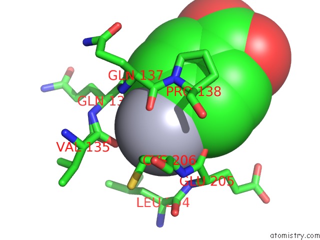
Mono view
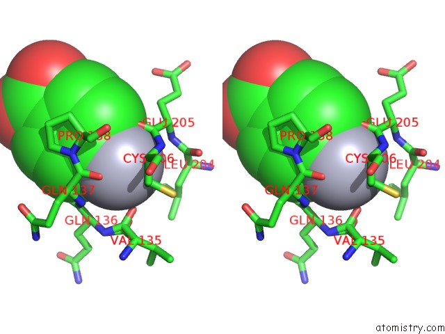
Stereo pair view
Mono view
Stereo pair view
A full contact list of Mercury with other atoms in the Hg binding
site number 1 of Carbonic Anhydrase II in Complex with N-[(2E)-3,4-Dihydroquinazolin- 2(1H)-Ylidene]Sulfuric Diamide within 5.0Å range:
|
Reference:
A.Di Pizio,
J.Schulze Wischeler,
M.Haake,
A.Heine,
C.Supuran,
G.Klebe.
High Resolution Crystal Structures of Carbonic Anhydrase II in Complex with Novel Sulfamide Binders To Be Published.
Page generated: Fri Aug 8 10:26:58 2025
Last articles
I in 4PVGI in 4PGC
I in 4PNS
I in 4P4Z
I in 4P1E
I in 4P4X
I in 4P4Y
I in 4P4W
I in 4OA5
I in 4OWA