Mercury »
PDB 4ihd-4q7w »
4kr6 »
Mercury in PDB 4kr6: Crystal Structure of A 4-Thiouridine Synthetase - Rna Complex
Enzymatic activity of Crystal Structure of A 4-Thiouridine Synthetase - Rna Complex
All present enzymatic activity of Crystal Structure of A 4-Thiouridine Synthetase - Rna Complex:
2.8.1.4;
2.8.1.4;
Protein crystallography data
The structure of Crystal Structure of A 4-Thiouridine Synthetase - Rna Complex, PDB code: 4kr6
was solved by
P.Neumann,
R.Ficner,
K.Lakomek,
with X-Ray Crystallography technique. A brief refinement statistics is given in the table below:
| Resolution Low / High (Å) | 30.00 / 2.85 |
| Space group | P 21 21 21 |
| Cell size a, b, c (Å), α, β, γ (°) | 100.652, 110.325, 119.313, 90.00, 90.00, 90.00 |
| R / Rfree (%) | 18.6 / 22.8 |
Mercury Binding Sites:
The binding sites of Mercury atom in the Crystal Structure of A 4-Thiouridine Synthetase - Rna Complex
(pdb code 4kr6). This binding sites where shown within
5.0 Angstroms radius around Mercury atom.
In total 4 binding sites of Mercury where determined in the Crystal Structure of A 4-Thiouridine Synthetase - Rna Complex, PDB code: 4kr6:
Jump to Mercury binding site number: 1; 2; 3; 4;
In total 4 binding sites of Mercury where determined in the Crystal Structure of A 4-Thiouridine Synthetase - Rna Complex, PDB code: 4kr6:
Jump to Mercury binding site number: 1; 2; 3; 4;
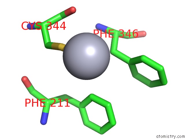
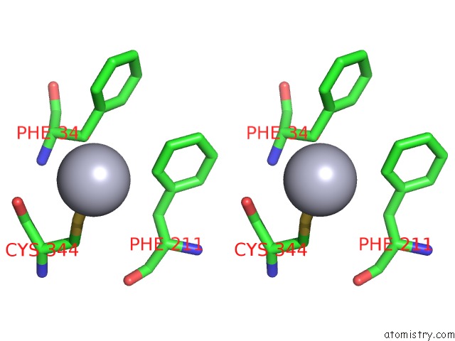
Mercury binding site 1 out of 4 in 4kr6
Go back to
Mercury binding site 1 out
of 4 in the Crystal Structure of A 4-Thiouridine Synthetase - Rna Complex
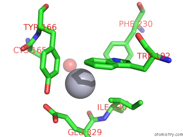
Mono view
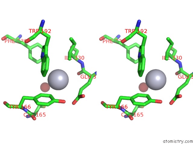
Stereo pair view
Mono view
Stereo pair view
A full contact list of Mercury with other atoms in the Hg binding
site number 1 of Crystal Structure of A 4-Thiouridine Synthetase - Rna Complex within 5.0Å range:
|
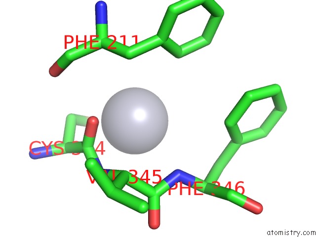
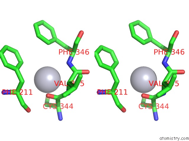
Mercury binding site 2 out of 4 in 4kr6
Go back to
Mercury binding site 2 out
of 4 in the Crystal Structure of A 4-Thiouridine Synthetase - Rna Complex
Mono view
Stereo pair view
Mono view
Stereo pair view
A full contact list of Mercury with other atoms in the Hg binding
site number 2 of Crystal Structure of A 4-Thiouridine Synthetase - Rna Complex within 5.0Å range:
|
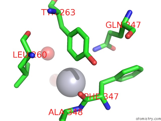
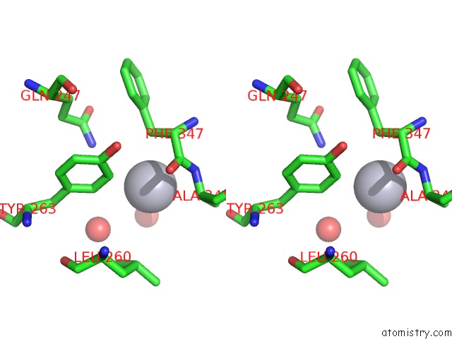
Mercury binding site 3 out of 4 in 4kr6
Go back to
Mercury binding site 3 out
of 4 in the Crystal Structure of A 4-Thiouridine Synthetase - Rna Complex
Mono view
Stereo pair view
Mono view
Stereo pair view
A full contact list of Mercury with other atoms in the Hg binding
site number 3 of Crystal Structure of A 4-Thiouridine Synthetase - Rna Complex within 5.0Å range:
|
Mercury binding site 4 out of 4 in 4kr6
Go back to
Mercury binding site 4 out
of 4 in the Crystal Structure of A 4-Thiouridine Synthetase - Rna Complex
Mono view
Stereo pair view
Mono view
Stereo pair view
A full contact list of Mercury with other atoms in the Hg binding
site number 4 of Crystal Structure of A 4-Thiouridine Synthetase - Rna Complex within 5.0Å range:
|
Reference:
P.Neumann,
K.Lakomek,
P.T.Naumann,
W.M.Erwin,
C.T.Lauhon,
R.Ficner.
Crystal Structure of A 4-Thiouridine Synthetase-Rna Complex Reveals Specificity of Trna U8 Modification. Nucleic Acids Res. V. 42 6673 2014.
ISSN: ISSN 0305-1048
PubMed: 24705700
DOI: 10.1093/NAR/GKU249
Page generated: Fri Aug 8 10:29:53 2025
ISSN: ISSN 0305-1048
PubMed: 24705700
DOI: 10.1093/NAR/GKU249
Last articles
I in 2X76I in 2XN6
I in 2X5X
I in 2XH1
I in 2XB5
I in 2WQ2
I in 2WPO
I in 2WKO
I in 2WAB
I in 2VMA