Atomistry » Mercury » PDB 4ihd-4q7w » 4mko
Atomistry »
  Mercury »
    PDB 4ihd-4q7w »
      4mko »

Mercury in PDB 4mko: Crystal Structure of the Monomeric, Cleaved Form of the Pore-Forming Toxin Monalysin

Protein crystallography data

The structure of Crystal Structure of the Monomeric, Cleaved Form of the Pore-Forming Toxin Monalysin, PDB code: 4mko was solved by P.Leone, A.Roussel, with X-Ray Crystallography technique. A brief refinement statistics is given in the table below:

Resolution Low / High (Å) 29.78 / 1.70
Space group P 21 21 21
Cell size a, b, c (Å), α, β, γ (°) 64.844, 117.649, 150.584, 90.00, 90.00, 90.00
R / Rfree (%) 17.9 / 20.5

Other elements in 4mko:

The structure of Crystal Structure of the Monomeric, Cleaved Form of the Pore-Forming Toxin Monalysin also contains other interesting chemical elements:

Zinc (Zn) 10 atoms

Mercury Binding Sites:

Pages:

>>> Page 1 <<< Page 2, Binding sites: 11 - 12;

Binding sites:

The binding sites of Mercury atom in the Crystal Structure of the Monomeric, Cleaved Form of the Pore-Forming Toxin Monalysin (pdb code 4mko). This binding sites where shown within 5.0 Angstroms radius around Mercury atom.
In total 12 binding sites of Mercury where determined in the Crystal Structure of the Monomeric, Cleaved Form of the Pore-Forming Toxin Monalysin, PDB code: 4mko:
Jump to Mercury binding site number: 1; 2; 3; 4; 5; 6; 7; 8; 9; 10;

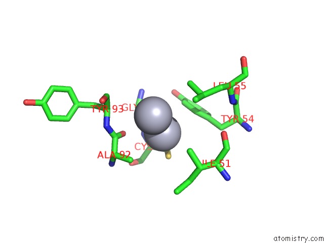
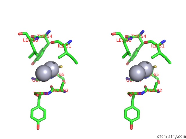
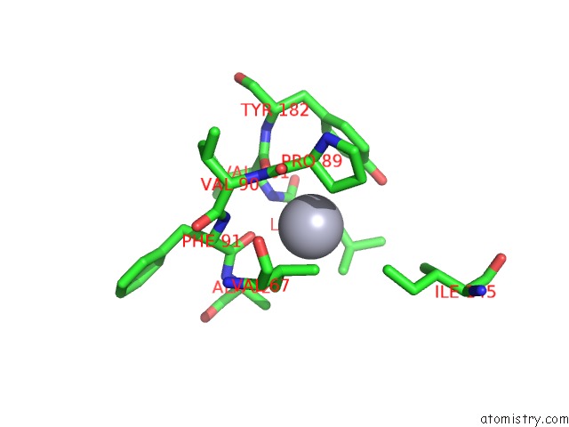
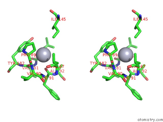
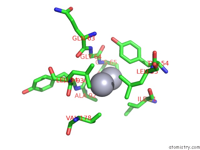
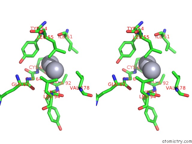
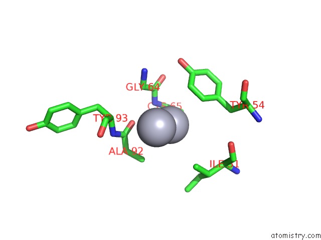
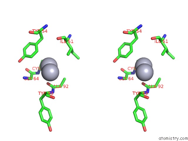
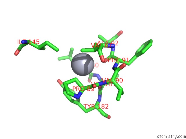
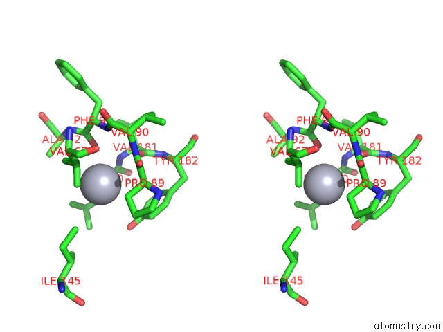
Mercury binding site 1 out of 12 in 4mko

Go back to Mercury Binding Sites List in 4mko
Mercury binding site 1 out of 12 in the Crystal Structure of the Monomeric, Cleaved Form of the Pore-Forming Toxin Monalysin


Mono view


Stereo pair view

A full contact list of Mercury with other atoms in the Hg binding site number 1 of Crystal Structure of the Monomeric, Cleaved Form of the Pore-Forming Toxin Monalysin within 5.0Å range:
probe atom residue distance (Å) B Occ
A:Hg1001

b:14.1
occ:0.45
HG A:HG1001 0.0 14.1 0.5
HG A:HG1001 2.2 17.5 0.4
SG A:CYS65 2.4 13.6 1.0
N A:CYS65 3.2 7.8 1.0
O A:ALA92 3.3 8.4 1.0
CB A:CYS65 3.3 9.1 1.0
CD2 A:TYR54 3.5 14.4 1.0
CE2 A:TYR54 3.6 16.0 1.0
C A:GLY64 3.7 11.0 1.0
CB A:ALA92 3.8 5.0 1.0
CA A:GLY64 3.8 9.7 1.0
CA A:CYS65 3.8 8.2 1.0
C A:ALA92 3.9 9.0 1.0
CG2 A:ILE51 4.1 12.7 1.0
CA A:ALA92 4.4 4.1 1.0
O A:GLY64 4.5 8.9 1.0
O A:CYS65 4.5 8.5 1.0
CG A:TYR54 4.5 13.0 1.0
C A:CYS65 4.6 10.0 1.0
CZ A:TYR54 4.6 22.6 1.0
N A:TYR93 4.7 5.0 1.0
N A:GLY64 4.8 9.9 1.0
CG1 A:ILE51 4.9 12.2 1.0
N A:ALA92 4.9 4.1 1.0
CB A:ILE51 5.0 12.0 1.0
CA A:TYR93 5.0 5.2 1.0
CD2 A:LEU55 5.0 11.8 1.0

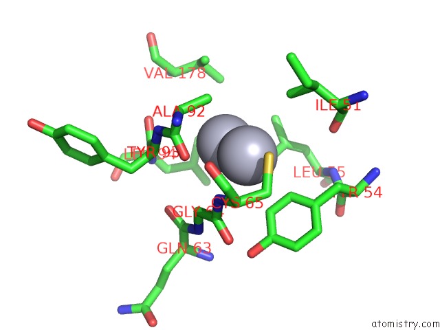
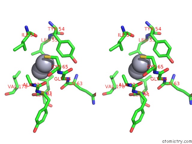
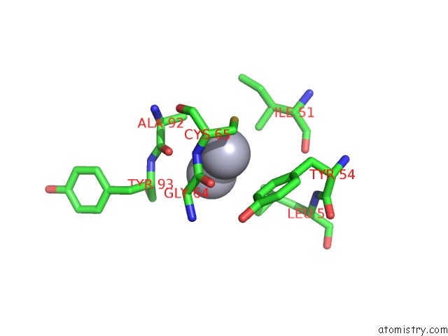
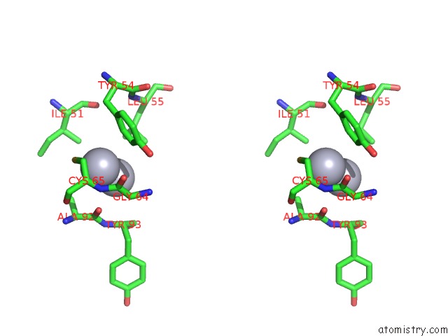
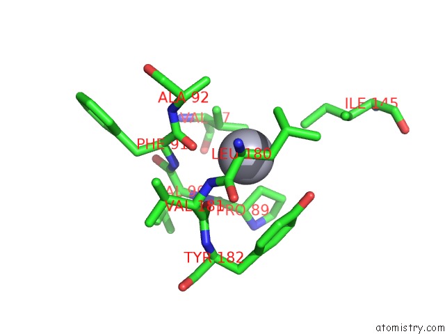
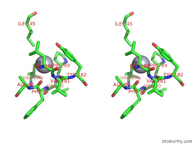
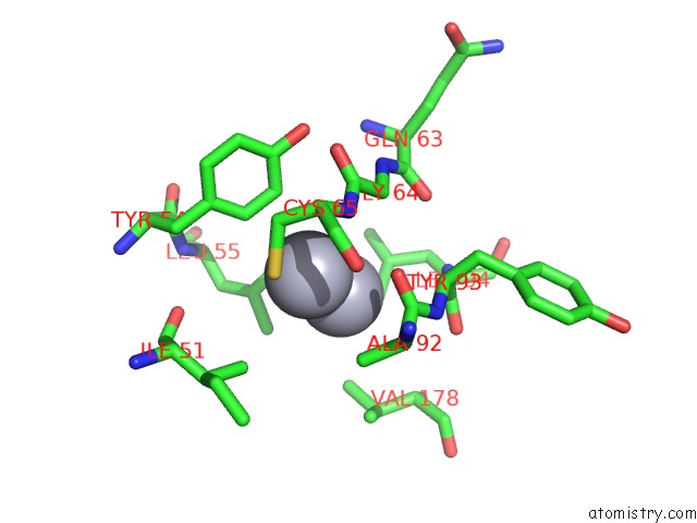
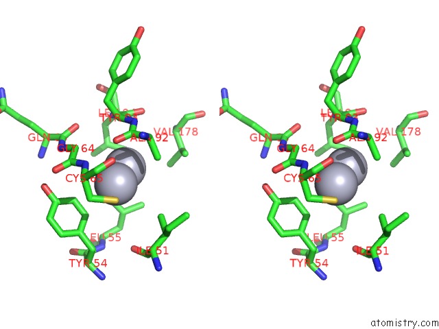
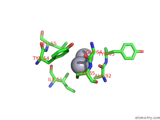
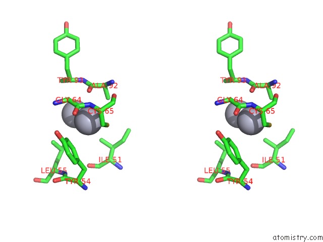
Mercury binding site 2 out of 12 in 4mko

Go back to Mercury Binding Sites List in 4mko
Mercury binding site 2 out of 12 in the Crystal Structure of the Monomeric, Cleaved Form of the Pore-Forming Toxin Monalysin


Mono view


Stereo pair view

A full contact list of Mercury with other atoms in the Hg binding site number 2 of Crystal Structure of the Monomeric, Cleaved Form of the Pore-Forming Toxin Monalysin within 5.0Å range:
probe atom residue distance (Å) B Occ
A:Hg1001

b:17.5
occ:0.40
HG A:HG1001 0.0 17.5 0.4
HG A:HG1001 2.2 14.1 0.5
O A:ALA92 3.5 8.4 1.0
C A:ALA92 3.7 9.0 1.0
CA A:TYR93 3.8 5.2 1.0
N A:TYR93 3.9 5.0 1.0
CD2 A:LEU55 4.0 11.8 1.0
CB A:ALA92 4.0 5.0 1.0
C A:TYR93 4.1 8.8 1.0
CA A:GLY64 4.1 9.7 1.0
CE2 A:TYR54 4.4 16.0 1.0
CG2 A:ILE51 4.4 12.7 1.0
CG A:LEU55 4.4 10.9 1.0
CD1 A:LEU55 4.4 11.2 1.0
O A:TYR93 4.4 8.7 1.0
N A:LEU94 4.5 6.1 1.0
CG A:LEU94 4.5 9.4 1.0
CD2 A:TYR54 4.5 14.4 1.0
CA A:ALA92 4.5 4.1 1.0
SG A:CYS65 4.5 13.6 1.0
CD2 A:LEU94 4.6 8.9 1.0
N A:CYS65 4.6 7.8 1.0
O A:GLN63 4.7 12.8 1.0
CG1 A:VAL178 4.7 7.6 1.0
C A:GLY64 4.7 11.0 1.0
N A:GLY64 4.8 9.9 1.0
CG2 A:VAL178 4.9 8.2 1.0

Mercury binding site 3 out of 12 in 4mko

Go back to Mercury Binding Sites List in 4mko
Mercury binding site 3 out of 12 in the Crystal Structure of the Monomeric, Cleaved Form of the Pore-Forming Toxin Monalysin


Mono view


Stereo pair view

A full contact list of Mercury with other atoms in the Hg binding site number 3 of Crystal Structure of the Monomeric, Cleaved Form of the Pore-Forming Toxin Monalysin within 5.0Å range:
probe atom residue distance (Å) B Occ
A:Hg1002

b:14.2
occ:0.09
O A:PHE91 3.0 5.8 1.0
O A:VAL181 3.4 4.5 1.0
CB A:PRO89 3.5 5.7 1.0
N A:VAL181 3.5 4.4 1.0
CB A:LEU180 3.6 4.6 1.0
CA A:LEU180 3.7 4.5 1.0
CD2 A:LEU180 3.7 10.7 1.0
CG2 A:VAL67 3.8 9.8 1.0
C A:LEU180 3.9 7.2 1.0
C A:PHE91 4.0 6.8 1.0
CB A:VAL67 4.1 9.9 1.0
CG1 A:VAL67 4.1 9.1 1.0
CA A:PRO89 4.2 4.0 1.0
C A:VAL181 4.2 6.9 1.0
CG A:LEU180 4.3 8.3 1.0
N A:PHE91 4.3 5.0 1.0
CZ A:TYR182 4.3 9.6 1.0
N A:VAL90 4.3 3.1 1.0
OH A:TYR182 4.4 10.4 1.0
CA A:VAL181 4.4 3.0 1.0
CD1 A:ILE145 4.5 16.5 1.0
CE2 A:TYR182 4.5 7.0 1.0
C A:PRO89 4.6 5.8 1.0
CE1 A:TYR182 4.7 5.4 1.0
CB A:ALA92 4.7 5.0 1.0
CG A:PRO89 4.8 9.3 1.0
CA A:PHE91 4.8 4.5 1.0
N A:ALA92 4.8 4.1 1.0
CA A:ALA92 4.9 4.1 1.0
O A:LEU180 5.0 7.2 1.0
CD2 A:TYR182 5.0 5.7 1.0

Mercury binding site 4 out of 12 in 4mko

Go back to Mercury Binding Sites List in 4mko
Mercury binding site 4 out of 12 in the Crystal Structure of the Monomeric, Cleaved Form of the Pore-Forming Toxin Monalysin


Mono view


Stereo pair view

A full contact list of Mercury with other atoms in the Hg binding site number 4 of Crystal Structure of the Monomeric, Cleaved Form of the Pore-Forming Toxin Monalysin within 5.0Å range:
probe atom residue distance (Å) B Occ
B:Hg1001

b:13.6
occ:0.43
HG B:HG1001 0.0 13.6 0.4
HG B:HG1001 2.2 18.0 0.4
SG B:CYS65 2.4 13.3 1.0
N B:CYS65 3.2 8.4 1.0
CB B:CYS65 3.3 8.9 1.0
O B:ALA92 3.4 7.0 1.0
CD2 B:TYR54 3.5 15.0 1.0
CE2 B:TYR54 3.6 17.3 1.0
C B:GLY64 3.7 10.8 1.0
CB B:ALA92 3.8 4.3 1.0
CA B:CYS65 3.8 8.4 1.0
CA B:GLY64 3.9 9.7 1.0
C B:ALA92 3.9 8.4 1.0
CG2 B:ILE51 4.1 10.7 1.0
CA B:ALA92 4.4 3.7 1.0
O B:CYS65 4.4 6.5 1.0
O B:GLY64 4.5 8.7 1.0
CG B:TYR54 4.5 13.4 1.0
CZ B:TYR54 4.6 24.6 1.0
C B:CYS65 4.6 9.4 1.0
N B:TYR93 4.7 4.7 1.0
N B:GLY64 4.8 9.8 1.0
CD2 B:LEU55 4.8 13.3 1.0
N B:ALA92 4.9 4.1 1.0
CA B:TYR93 4.9 5.3 1.0
CG1 B:ILE51 5.0 8.8 1.0

Mercury binding site 5 out of 12 in 4mko

Go back to Mercury Binding Sites List in 4mko
Mercury binding site 5 out of 12 in the Crystal Structure of the Monomeric, Cleaved Form of the Pore-Forming Toxin Monalysin


Mono view


Stereo pair view

A full contact list of Mercury with other atoms in the Hg binding site number 5 of Crystal Structure of the Monomeric, Cleaved Form of the Pore-Forming Toxin Monalysin within 5.0Å range:
probe atom residue distance (Å) B Occ
B:Hg1001

b:18.0
occ:0.40
HG B:HG1001 0.0 18.0 0.4
HG B:HG1001 2.2 13.6 0.4
O B:ALA92 3.6 7.0 1.0
C B:ALA92 3.7 8.4 1.0
CA B:TYR93 3.8 5.3 1.0
N B:TYR93 3.8 4.7 1.0
CD2 B:LEU55 3.8 13.3 1.0
CB B:ALA92 3.9 4.3 1.0
C B:TYR93 4.0 8.2 1.0
CA B:GLY64 4.2 9.7 1.0
N B:LEU94 4.4 6.5 1.0
CG B:LEU55 4.4 12.1 1.0
CE2 B:TYR54 4.4 17.3 1.0
CG2 B:ILE51 4.4 10.7 1.0
CA B:ALA92 4.4 3.7 1.0
O B:TYR93 4.4 7.1 1.0
CD1 B:LEU55 4.5 12.7 1.0
SG B:CYS65 4.6 13.3 1.0
CG B:LEU94 4.6 9.4 1.0
CD2 B:TYR54 4.6 15.0 1.0
CD2 B:LEU94 4.6 11.9 1.0
N B:CYS65 4.7 8.4 1.0
CG1 B:VAL178 4.7 6.4 1.0
CG2 B:VAL178 4.7 6.2 1.0
O B:GLN63 4.8 12.9 1.0
C B:GLY64 4.8 10.8 1.0
N B:GLY64 4.9 9.8 1.0

Mercury binding site 6 out of 12 in 4mko

Go back to Mercury Binding Sites List in 4mko
Mercury binding site 6 out of 12 in the Crystal Structure of the Monomeric, Cleaved Form of the Pore-Forming Toxin Monalysin


Mono view


Stereo pair view

A full contact list of Mercury with other atoms in the Hg binding site number 6 of Crystal Structure of the Monomeric, Cleaved Form of the Pore-Forming Toxin Monalysin within 5.0Å range:
probe atom residue distance (Å) B Occ
B:Hg1002

b:15.4
occ:0.07
O B:PHE91 3.1 6.5 1.0
CB B:PRO89 3.4 5.5 1.0
O B:VAL181 3.4 6.1 1.0
N B:VAL181 3.5 3.8 1.0
CB B:LEU180 3.5 3.8 1.0
CA B:LEU180 3.7 3.9 1.0
CD2 B:LEU180 3.7 7.4 1.0
CG2 B:VAL67 3.7 9.7 1.0
C B:LEU180 3.9 6.7 1.0
C B:PHE91 4.0 6.9 1.0
CB B:VAL67 4.0 9.5 1.0
CG1 B:VAL67 4.1 9.5 1.0
CA B:PRO89 4.1 4.3 1.0
C B:VAL181 4.2 6.1 1.0
CG B:LEU180 4.2 7.0 1.0
CZ B:TYR182 4.2 9.3 1.0
N B:PHE91 4.2 4.3 1.0
CD1 B:ILE145 4.3 11.3 1.0
N B:VAL90 4.3 3.4 1.0
OH B:TYR182 4.3 8.5 1.0
CE2 B:TYR182 4.4 7.0 1.0
CA B:VAL181 4.5 3.0 1.0
C B:PRO89 4.6 7.0 1.0
CE1 B:TYR182 4.6 6.4 1.0
CG B:PRO89 4.7 9.2 1.0
CA B:PHE91 4.7 4.3 1.0
CB B:ALA92 4.8 4.3 1.0
N B:ALA92 4.8 4.1 1.0
O B:LEU180 4.9 6.6 1.0
CD2 B:TYR182 4.9 5.2 1.0
CA B:ALA92 5.0 3.7 1.0

Mercury binding site 7 out of 12 in 4mko

Go back to Mercury Binding Sites List in 4mko
Mercury binding site 7 out of 12 in the Crystal Structure of the Monomeric, Cleaved Form of the Pore-Forming Toxin Monalysin


Mono view


Stereo pair view

A full contact list of Mercury with other atoms in the Hg binding site number 7 of Crystal Structure of the Monomeric, Cleaved Form of the Pore-Forming Toxin Monalysin within 5.0Å range:
probe atom residue distance (Å) B Occ
C:Hg1001

b:14.8
occ:0.44
HG C:HG1001 0.0 14.8 0.4
HG C:HG1001 2.2 16.8 0.4
SG C:CYS65 2.4 13.8 1.0
N C:CYS65 3.2 9.0 1.0
O C:ALA92 3.3 9.8 1.0
CB C:CYS65 3.3 9.5 1.0
CD2 C:TYR54 3.5 14.7 1.0
CE2 C:TYR54 3.6 16.3 1.0
C C:GLY64 3.7 11.4 1.0
CB C:ALA92 3.8 5.9 1.0
CA C:CYS65 3.8 9.0 1.0
C C:ALA92 3.8 10.8 1.0
CA C:GLY64 3.9 9.4 1.0
CG2 C:ILE51 4.2 12.6 1.0
O C:CYS65 4.3 7.9 1.0
CA C:ALA92 4.4 5.0 1.0
CG C:TYR54 4.5 12.6 1.0
O C:GLY64 4.5 10.1 1.0
C C:CYS65 4.5 9.8 1.0
CZ C:TYR54 4.6 24.3 1.0
N C:TYR93 4.7 6.5 1.0
N C:GLY64 4.8 10.1 1.0
CA C:TYR93 4.9 7.0 1.0
N C:ALA92 4.9 4.0 1.0

Mercury binding site 8 out of 12 in 4mko

Go back to Mercury Binding Sites List in 4mko
Mercury binding site 8 out of 12 in the Crystal Structure of the Monomeric, Cleaved Form of the Pore-Forming Toxin Monalysin


Mono view


Stereo pair view

A full contact list of Mercury with other atoms in the Hg binding site number 8 of Crystal Structure of the Monomeric, Cleaved Form of the Pore-Forming Toxin Monalysin within 5.0Å range:
probe atom residue distance (Å) B Occ
C:Hg1001

b:16.8
occ:0.38
HG C:HG1001 0.0 16.8 0.4
HG C:HG1001 2.2 14.8 0.4
O C:ALA92 3.5 9.8 1.0
C C:ALA92 3.6 10.8 1.0
CA C:TYR93 3.8 7.0 1.0
N C:TYR93 3.8 6.5 1.0
CD2 C:LEU55 4.0 14.0 1.0
CB C:ALA92 4.0 5.9 1.0
C C:TYR93 4.0 10.0 1.0
CA C:GLY64 4.2 9.4 1.0
CD1 C:LEU55 4.3 13.1 1.0
CG C:LEU55 4.4 11.8 1.0
N C:LEU94 4.4 8.1 1.0
CE2 C:TYR54 4.4 16.3 1.0
CA C:ALA92 4.5 5.0 1.0
CG C:LEU94 4.5 11.9 1.0
CG2 C:ILE51 4.5 12.6 1.0
CD2 C:TYR54 4.5 14.7 1.0
O C:TYR93 4.5 8.6 1.0
SG C:CYS65 4.5 13.8 1.0
CD2 C:LEU94 4.6 13.6 1.0
N C:CYS65 4.7 9.0 1.0
CG1 C:VAL178 4.7 8.2 1.0
O C:GLN63 4.8 13.8 1.0
CG2 C:VAL178 4.8 8.5 1.0
C C:GLY64 4.8 11.4 1.0
N C:GLY64 4.9 10.1 1.0

Mercury binding site 9 out of 12 in 4mko

Go back to Mercury Binding Sites List in 4mko
Mercury binding site 9 out of 12 in the Crystal Structure of the Monomeric, Cleaved Form of the Pore-Forming Toxin Monalysin


Mono view


Stereo pair view

A full contact list of Mercury with other atoms in the Hg binding site number 9 of Crystal Structure of the Monomeric, Cleaved Form of the Pore-Forming Toxin Monalysin within 5.0Å range:
probe atom residue distance (Å) B Occ
C:Hg1002

b:17.5
occ:0.08
O C:PHE91 3.1 7.2 1.0
CB C:PRO89 3.5 6.1 1.0
O C:VAL181 3.6 6.2 1.0
CB C:LEU180 3.6 3.5 1.0
CD2 C:LEU180 3.6 8.4 1.0
N C:VAL181 3.6 6.0 1.0
CG2 C:VAL67 3.7 9.4 1.0
CA C:LEU180 3.8 3.8 1.0
C C:LEU180 4.0 8.3 1.0
C C:PHE91 4.1 6.7 1.0
CB C:VAL67 4.1 9.1 1.0
CG1 C:VAL67 4.1 8.5 1.0
CG C:LEU180 4.2 7.5 1.0
CA C:PRO89 4.2 4.3 1.0
CD1 C:ILE145 4.3 14.1 1.0
CZ C:TYR182 4.3 7.8 1.0
C C:VAL181 4.3 7.7 1.0
OH C:TYR182 4.3 7.2 1.0
N C:PHE91 4.4 5.1 1.0
N C:VAL90 4.4 3.9 1.0
CE2 C:TYR182 4.5 6.5 1.0
CA C:VAL181 4.6 5.1 1.0
CE1 C:TYR182 4.7 6.8 1.0
C C:PRO89 4.7 7.1 1.0
CB C:ALA92 4.7 5.9 1.0
CG C:PRO89 4.7 9.8 1.0
N C:ALA92 4.8 4.0 1.0
CA C:PHE91 4.9 5.2 1.0
CA C:ALA92 5.0 5.0 1.0

Mercury binding site 10 out of 12 in 4mko

Go back to Mercury Binding Sites List in 4mko
Mercury binding site 10 out of 12 in the Crystal Structure of the Monomeric, Cleaved Form of the Pore-Forming Toxin Monalysin


Mono view


Stereo pair view

A full contact list of Mercury with other atoms in the Hg binding site number 10 of Crystal Structure of the Monomeric, Cleaved Form of the Pore-Forming Toxin Monalysin within 5.0Å range:
probe atom residue distance (Å) B Occ
D:Hg1001

b:15.9
occ:0.43
HG D:HG1001 0.0 15.9 0.4
HG D:HG1001 2.3 18.6 0.4
SG D:CYS65 2.3 14.9 1.0
N D:CYS65 3.3 6.3 1.0
O D:ALA92 3.3 8.9 1.0
CB D:CYS65 3.3 10.0 1.0
CD2 D:TYR54 3.6 18.7 1.0
CE2 D:TYR54 3.6 20.7 1.0
C D:GLY64 3.7 10.9 1.0
CB D:ALA92 3.8 5.3 1.0
CA D:CYS65 3.9 8.4 1.0
CA D:GLY64 3.9 9.1 1.0
C D:ALA92 3.9 9.0 1.0
CG2 D:ILE51 4.2 13.8 1.0
CA D:ALA92 4.4 4.9 1.0
O D:GLY64 4.4 10.6 1.0
O D:CYS65 4.5 8.4 1.0
C D:CYS65 4.6 11.0 1.0
CG D:TYR54 4.7 16.7 1.0
N D:TYR93 4.7 5.5 1.0
CZ D:TYR54 4.7 29.5 1.0
N D:GLY64 4.8 10.5 1.0
N D:ALA92 4.9 5.3 1.0
CA D:TYR93 5.0 4.5 1.0
CD2 D:LEU55 5.0 8.5 1.0

Reference:

P.Leone, A.Roussel. A Monalysin Toxin Hypothetical Pore Formation Mechanism Deduced From X-Ray and Cryoem Studies To Be Published.
Page generated: Sun Dec 13 19:11:27 2020

Last articles

Zn in 8WB0
Zn in 8WAX
Zn in 8WAU
Zn in 8WAZ
Zn in 8WAY
Zn in 8WAV
Zn in 8WAW
Zn in 8WAT
Zn in 8W7M
Zn in 8WD3
© Copyright 2008-2020 by atomistry.com
Home   |    Site Map   |    Copyright   |    Contact us   |    Privacy