Mercury »
PDB 4ihd-4q7w »
4q2e »
Mercury in PDB 4q2e: Crystal Structure of An Intramembrane Cdp-Dag Synthetase Central For Phospholipid Biosynthesis (S200C/S258C, Active Mutant)
Enzymatic activity of Crystal Structure of An Intramembrane Cdp-Dag Synthetase Central For Phospholipid Biosynthesis (S200C/S258C, Active Mutant)
All present enzymatic activity of Crystal Structure of An Intramembrane Cdp-Dag Synthetase Central For Phospholipid Biosynthesis (S200C/S258C, Active Mutant):
2.7.7.41;
2.7.7.41;
Protein crystallography data
The structure of Crystal Structure of An Intramembrane Cdp-Dag Synthetase Central For Phospholipid Biosynthesis (S200C/S258C, Active Mutant), PDB code: 4q2e
was solved by
X.Liu,
Y.Yin,
J.Wu,
Z.Liu,
with X-Ray Crystallography technique. A brief refinement statistics is given in the table below:
| Resolution Low / High (Å) | 45.00 / 3.40 |
| Space group | P 65 2 2 |
| Cell size a, b, c (Å), α, β, γ (°) | 142.080, 142.080, 198.373, 90.00, 90.00, 120.00 |
| R / Rfree (%) | 29.9 / 33.8 |
Other elements in 4q2e:
The structure of Crystal Structure of An Intramembrane Cdp-Dag Synthetase Central For Phospholipid Biosynthesis (S200C/S258C, Active Mutant) also contains other interesting chemical elements:
| Magnesium | (Mg) | 2 atoms |
| Potassium | (K) | 2 atoms |
Mercury Binding Sites:
The binding sites of Mercury atom in the Crystal Structure of An Intramembrane Cdp-Dag Synthetase Central For Phospholipid Biosynthesis (S200C/S258C, Active Mutant)
(pdb code 4q2e). This binding sites where shown within
5.0 Angstroms radius around Mercury atom.
In total 6 binding sites of Mercury where determined in the Crystal Structure of An Intramembrane Cdp-Dag Synthetase Central For Phospholipid Biosynthesis (S200C/S258C, Active Mutant), PDB code: 4q2e:
Jump to Mercury binding site number: 1; 2; 3; 4; 5; 6;
In total 6 binding sites of Mercury where determined in the Crystal Structure of An Intramembrane Cdp-Dag Synthetase Central For Phospholipid Biosynthesis (S200C/S258C, Active Mutant), PDB code: 4q2e:
Jump to Mercury binding site number: 1; 2; 3; 4; 5; 6;
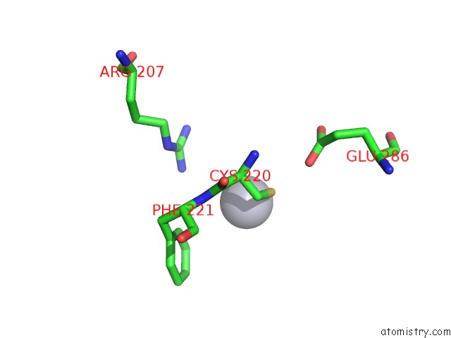
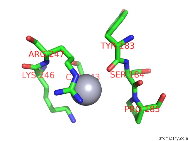
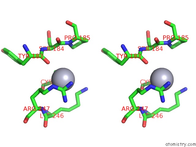
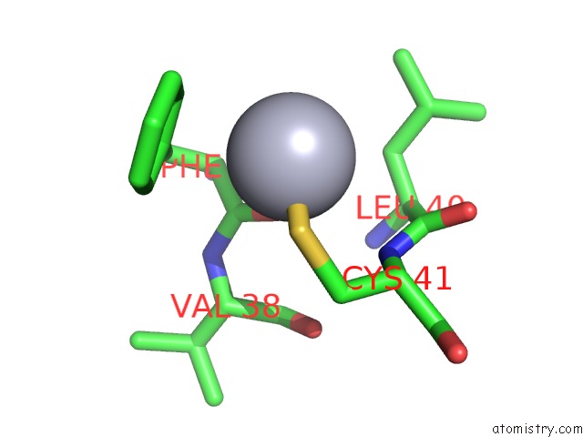
Mercury binding site 1 out of 6 in 4q2e
Go back to
Mercury binding site 1 out
of 6 in the Crystal Structure of An Intramembrane Cdp-Dag Synthetase Central For Phospholipid Biosynthesis (S200C/S258C, Active Mutant)
Mono view
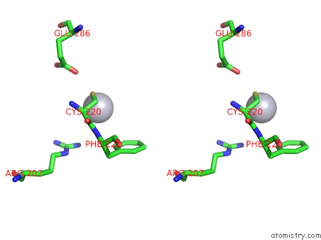
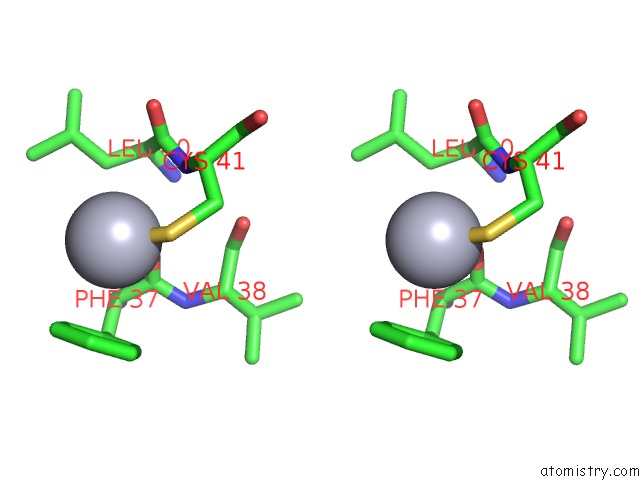
Stereo pair view
Mono view
Stereo pair view
A full contact list of Mercury with other atoms in the Hg binding
site number 1 of Crystal Structure of An Intramembrane Cdp-Dag Synthetase Central For Phospholipid Biosynthesis (S200C/S258C, Active Mutant) within 5.0Å range:
|
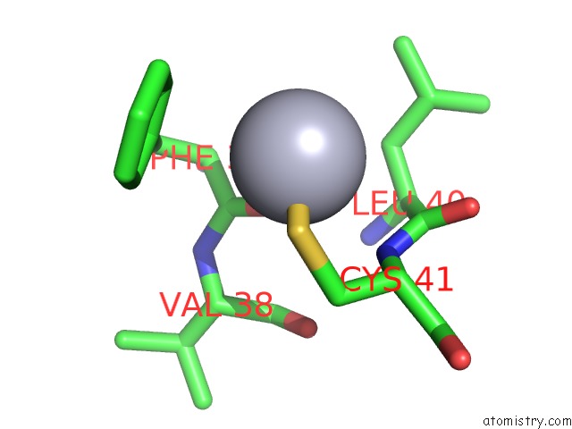
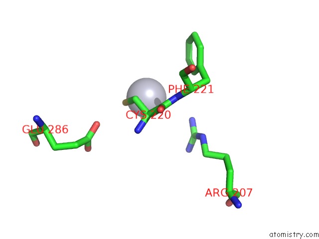
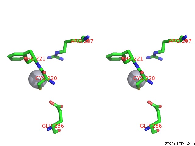
Mercury binding site 2 out of 6 in 4q2e
Go back to
Mercury binding site 2 out
of 6 in the Crystal Structure of An Intramembrane Cdp-Dag Synthetase Central For Phospholipid Biosynthesis (S200C/S258C, Active Mutant)
Mono view
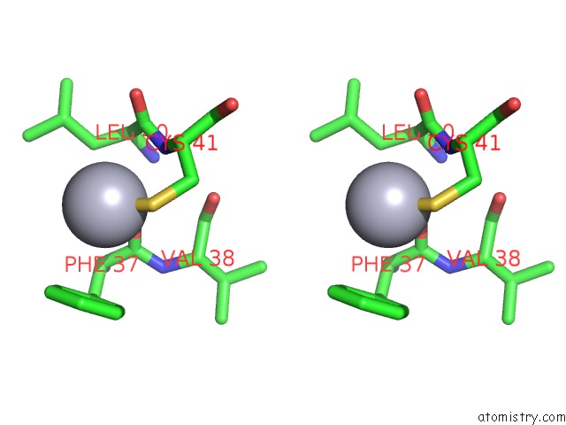
Stereo pair view
Mono view
Stereo pair view
A full contact list of Mercury with other atoms in the Hg binding
site number 2 of Crystal Structure of An Intramembrane Cdp-Dag Synthetase Central For Phospholipid Biosynthesis (S200C/S258C, Active Mutant) within 5.0Å range:
|
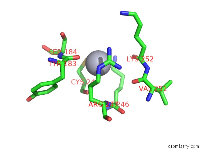
Mercury binding site 3 out of 6 in 4q2e
Go back to
Mercury binding site 3 out
of 6 in the Crystal Structure of An Intramembrane Cdp-Dag Synthetase Central For Phospholipid Biosynthesis (S200C/S258C, Active Mutant)
Mono view
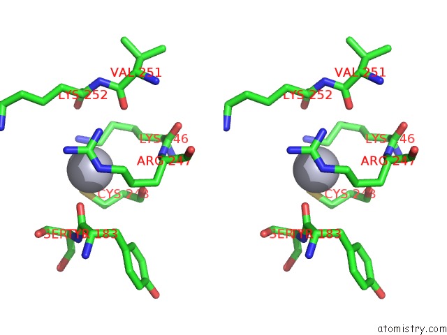
Stereo pair view
Mono view
Stereo pair view
A full contact list of Mercury with other atoms in the Hg binding
site number 3 of Crystal Structure of An Intramembrane Cdp-Dag Synthetase Central For Phospholipid Biosynthesis (S200C/S258C, Active Mutant) within 5.0Å range:
|
Mercury binding site 4 out of 6 in 4q2e
Go back to
Mercury binding site 4 out
of 6 in the Crystal Structure of An Intramembrane Cdp-Dag Synthetase Central For Phospholipid Biosynthesis (S200C/S258C, Active Mutant)
Mono view
Stereo pair view
Mono view
Stereo pair view
A full contact list of Mercury with other atoms in the Hg binding
site number 4 of Crystal Structure of An Intramembrane Cdp-Dag Synthetase Central For Phospholipid Biosynthesis (S200C/S258C, Active Mutant) within 5.0Å range:
|
Mercury binding site 5 out of 6 in 4q2e
Go back to
Mercury binding site 5 out
of 6 in the Crystal Structure of An Intramembrane Cdp-Dag Synthetase Central For Phospholipid Biosynthesis (S200C/S258C, Active Mutant)
Mono view
Stereo pair view
Mono view
Stereo pair view
A full contact list of Mercury with other atoms in the Hg binding
site number 5 of Crystal Structure of An Intramembrane Cdp-Dag Synthetase Central For Phospholipid Biosynthesis (S200C/S258C, Active Mutant) within 5.0Å range:
|
Mercury binding site 6 out of 6 in 4q2e
Go back to
Mercury binding site 6 out
of 6 in the Crystal Structure of An Intramembrane Cdp-Dag Synthetase Central For Phospholipid Biosynthesis (S200C/S258C, Active Mutant)
Mono view
Stereo pair view
Mono view
Stereo pair view
A full contact list of Mercury with other atoms in the Hg binding
site number 6 of Crystal Structure of An Intramembrane Cdp-Dag Synthetase Central For Phospholipid Biosynthesis (S200C/S258C, Active Mutant) within 5.0Å range:
|
Reference:
X.Liu,
Y.Yin,
J.Wu,
Z.Liu.
Structure and Mechanism of An Intramembrane Liponucleotide Synthetase Central For Phospholipid Biosynthesis Nat Commun V. 5 4244 2014.
ISSN: ESSN 2041-1723
PubMed: 24968740
DOI: 10.1038/NCOMMS5244
Page generated: Sun Aug 11 05:07:49 2024
ISSN: ESSN 2041-1723
PubMed: 24968740
DOI: 10.1038/NCOMMS5244
Last articles
Zn in 9MJ5Zn in 9HNW
Zn in 9G0L
Zn in 9FNE
Zn in 9DZN
Zn in 9E0I
Zn in 9D32
Zn in 9DAK
Zn in 8ZXC
Zn in 8ZUF