Mercury »
PDB 4q81-5c9l »
4ri2 »
Mercury in PDB 4ri2: Crystal Structure of the Photoprotective Protein Psbs From Spinach
Protein crystallography data
The structure of Crystal Structure of the Photoprotective Protein Psbs From Spinach, PDB code: 4ri2
was solved by
M.Fan,
M.Li,
W.Chang,
with X-Ray Crystallography technique. A brief refinement statistics is given in the table below:
| Resolution Low / High (Å) | 29.80 / 2.35 |
| Space group | P 21 21 21 |
| Cell size a, b, c (Å), α, β, γ (°) | 71.999, 77.537, 93.184, 90.00, 90.00, 90.00 |
| R / Rfree (%) | 23 / 26.6 |
Other elements in 4ri2:
The structure of Crystal Structure of the Photoprotective Protein Psbs From Spinach also contains other interesting chemical elements:
| Magnesium | (Mg) | 1 atom |
Mercury Binding Sites:
The binding sites of Mercury atom in the Crystal Structure of the Photoprotective Protein Psbs From Spinach
(pdb code 4ri2). This binding sites where shown within
5.0 Angstroms radius around Mercury atom.
In total only one binding site of Mercury was determined in the Crystal Structure of the Photoprotective Protein Psbs From Spinach, PDB code: 4ri2:
In total only one binding site of Mercury was determined in the Crystal Structure of the Photoprotective Protein Psbs From Spinach, PDB code: 4ri2:
Mercury binding site 1 out of 1 in 4ri2
Go back to
Mercury binding site 1 out
of 1 in the Crystal Structure of the Photoprotective Protein Psbs From Spinach
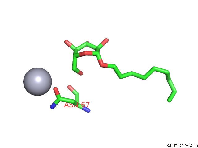
Mono view
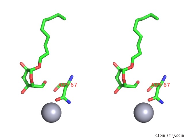
Stereo pair view
Mono view
Stereo pair view
A full contact list of Mercury with other atoms in the Hg binding
site number 1 of Crystal Structure of the Photoprotective Protein Psbs From Spinach within 5.0Å range:
|
Reference:
M.Fan,
M.Li,
Z.Liu,
P.Cao,
X.Pan,
H.Zhang,
X.Zhao,
J.Zhang,
W.Chang.
Crystal Structures of the Psbs Protein Essential For Photoprotection in Plants. Nat.Struct.Mol.Biol. V. 22 729 2015.
ISSN: ISSN 1545-9993
PubMed: 26258636
DOI: 10.1038/NSMB.3068
Page generated: Fri Aug 8 10:39:14 2025
ISSN: ISSN 1545-9993
PubMed: 26258636
DOI: 10.1038/NSMB.3068
Last articles
I in 3APBI in 3BAS
I in 3B56
I in 3B65
I in 3B2L
I in 3B2K
I in 3B2J
I in 3B2H
I in 3B2I
I in 3AEG