Mercury »
PDB 4q81-5c9l »
4ri3 »
Mercury in PDB 4ri3: Crystal Structure of Dccd-Modified Psbs From Spinach
Protein crystallography data
The structure of Crystal Structure of Dccd-Modified Psbs From Spinach, PDB code: 4ri3
was solved by
M.Fan,
M.Li,
W.Chang,
with X-Ray Crystallography technique. A brief refinement statistics is given in the table below:
| Resolution Low / High (Å) | 39.06 / 2.70 |
| Space group | P 21 21 21 |
| Cell size a, b, c (Å), α, β, γ (°) | 71.704, 77.697, 93.159, 90.00, 90.00, 90.00 |
| R / Rfree (%) | 23.2 / 28.4 |
Mercury Binding Sites:
The binding sites of Mercury atom in the Crystal Structure of Dccd-Modified Psbs From Spinach
(pdb code 4ri3). This binding sites where shown within
5.0 Angstroms radius around Mercury atom.
In total 7 binding sites of Mercury where determined in the Crystal Structure of Dccd-Modified Psbs From Spinach, PDB code: 4ri3:
Jump to Mercury binding site number: 1; 2; 3; 4; 5; 6; 7;
In total 7 binding sites of Mercury where determined in the Crystal Structure of Dccd-Modified Psbs From Spinach, PDB code: 4ri3:
Jump to Mercury binding site number: 1; 2; 3; 4; 5; 6; 7;
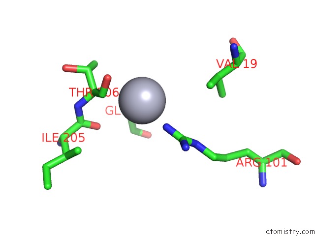
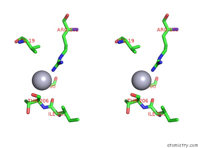
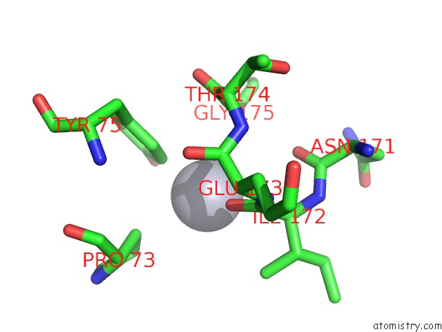
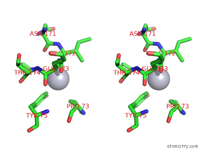
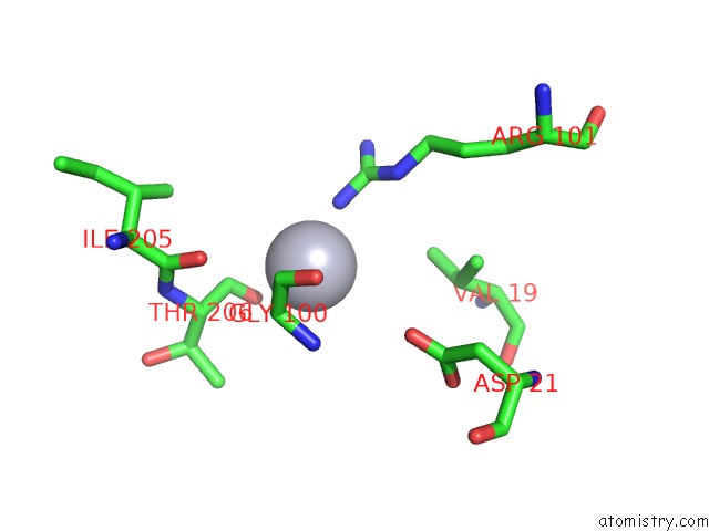
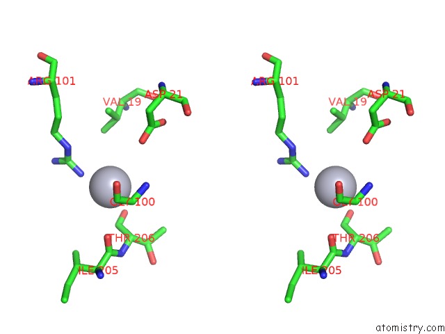
Mercury binding site 1 out of 7 in 4ri3
Go back to
Mercury binding site 1 out
of 7 in the Crystal Structure of Dccd-Modified Psbs From Spinach
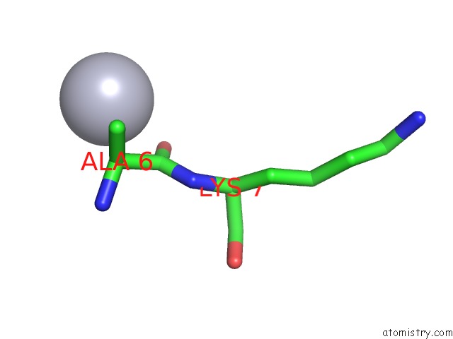
Mono view
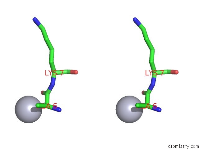
Stereo pair view
Mono view
Stereo pair view
A full contact list of Mercury with other atoms in the Hg binding
site number 1 of Crystal Structure of Dccd-Modified Psbs From Spinach within 5.0Å range:
|
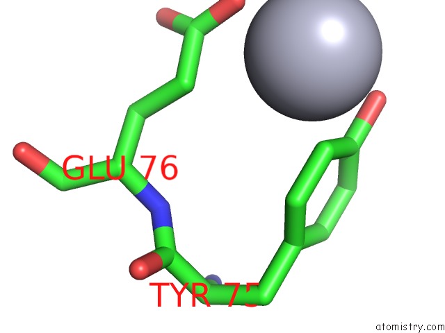
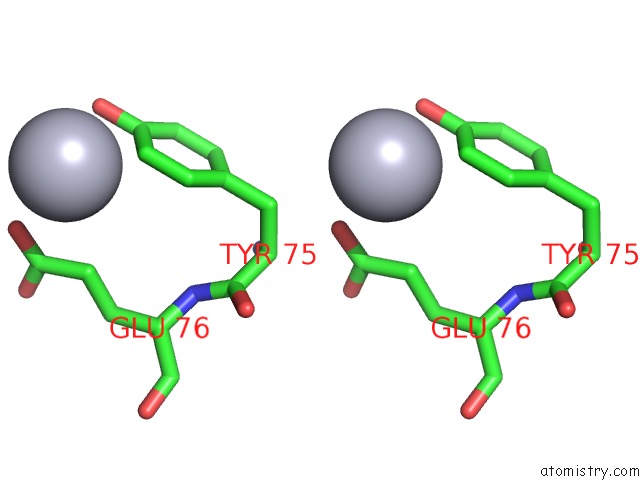
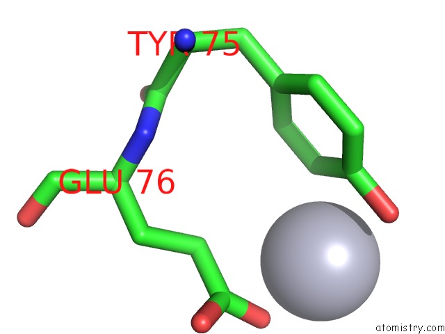
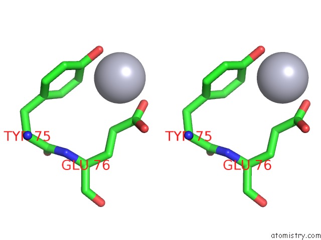
Mercury binding site 2 out of 7 in 4ri3
Go back to
Mercury binding site 2 out
of 7 in the Crystal Structure of Dccd-Modified Psbs From Spinach
Mono view
Stereo pair view
Mono view
Stereo pair view
A full contact list of Mercury with other atoms in the Hg binding
site number 2 of Crystal Structure of Dccd-Modified Psbs From Spinach within 5.0Å range:
|
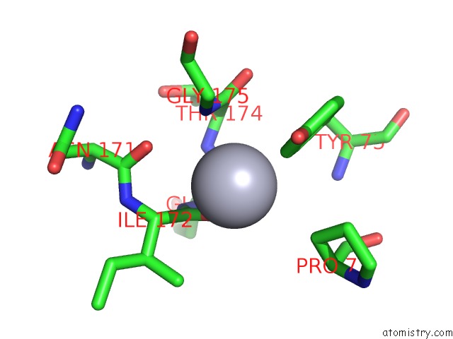
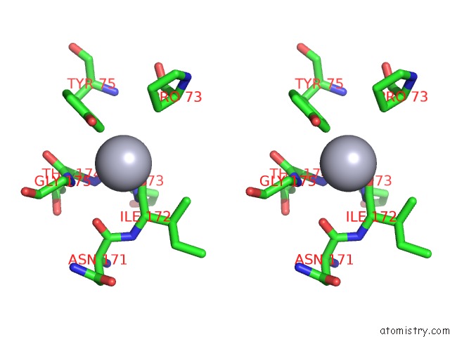
Mercury binding site 3 out of 7 in 4ri3
Go back to
Mercury binding site 3 out
of 7 in the Crystal Structure of Dccd-Modified Psbs From Spinach
Mono view
Stereo pair view
Mono view
Stereo pair view
A full contact list of Mercury with other atoms in the Hg binding
site number 3 of Crystal Structure of Dccd-Modified Psbs From Spinach within 5.0Å range:
|
Mercury binding site 4 out of 7 in 4ri3
Go back to
Mercury binding site 4 out
of 7 in the Crystal Structure of Dccd-Modified Psbs From Spinach
Mono view
Stereo pair view
Mono view
Stereo pair view
A full contact list of Mercury with other atoms in the Hg binding
site number 4 of Crystal Structure of Dccd-Modified Psbs From Spinach within 5.0Å range:
|
Mercury binding site 5 out of 7 in 4ri3
Go back to
Mercury binding site 5 out
of 7 in the Crystal Structure of Dccd-Modified Psbs From Spinach
Mono view
Stereo pair view
Mono view
Stereo pair view
A full contact list of Mercury with other atoms in the Hg binding
site number 5 of Crystal Structure of Dccd-Modified Psbs From Spinach within 5.0Å range:
|
Mercury binding site 6 out of 7 in 4ri3
Go back to
Mercury binding site 6 out
of 7 in the Crystal Structure of Dccd-Modified Psbs From Spinach
Mono view
Stereo pair view
Mono view
Stereo pair view
A full contact list of Mercury with other atoms in the Hg binding
site number 6 of Crystal Structure of Dccd-Modified Psbs From Spinach within 5.0Å range:
|
Mercury binding site 7 out of 7 in 4ri3
Go back to
Mercury binding site 7 out
of 7 in the Crystal Structure of Dccd-Modified Psbs From Spinach
Mono view
Stereo pair view
Mono view
Stereo pair view
A full contact list of Mercury with other atoms in the Hg binding
site number 7 of Crystal Structure of Dccd-Modified Psbs From Spinach within 5.0Å range:
|
Reference:
M.Fan,
M.Li,
Z.Liu,
P.Cao,
X.Pan,
H.Zhang,
X.Zhao,
J.Zhang,
W.Chang.
Crystal Structures of the Psbs Protein Essential For Photoprotection in Plants. Nat.Struct.Mol.Biol. V. 22 729 2015.
ISSN: ISSN 1545-9993
PubMed: 26258636
DOI: 10.1038/NSMB.3068
Page generated: Fri Aug 8 10:39:20 2025
ISSN: ISSN 1545-9993
PubMed: 26258636
DOI: 10.1038/NSMB.3068
Last articles
K in 2FRZK in 2FPQ
K in 2FPM
K in 2FPL
K in 2FPK
K in 2FPG
K in 2FP4
K in 2FEU
K in 2FFY
K in 2FDE