Mercury »
PDB 4q81-5c9l »
4uxm »
Mercury in PDB 4uxm: Crystal Structure of Struthiocalcin-1, A Different Crystal Form.
Protein crystallography data
The structure of Crystal Structure of Struthiocalcin-1, A Different Crystal Form., PDB code: 4uxm
was solved by
R.R.Ruiz-Arellano,
A.Moreno,
A.Romero,
with X-Ray Crystallography technique. A brief refinement statistics is given in the table below:
| Resolution Low / High (Å) | 44.173 / 1.50 |
| Space group | P 21 21 21 |
| Cell size a, b, c (Å), α, β, γ (°) | 32.670, 55.910, 72.060, 90.00, 90.00, 90.00 |
| R / Rfree (%) | 18.81 / 21.32 |
Other elements in 4uxm:
The structure of Crystal Structure of Struthiocalcin-1, A Different Crystal Form. also contains other interesting chemical elements:
| Chlorine | (Cl) | 1 atom |
Mercury Binding Sites:
The binding sites of Mercury atom in the Crystal Structure of Struthiocalcin-1, A Different Crystal Form.
(pdb code 4uxm). This binding sites where shown within
5.0 Angstroms radius around Mercury atom.
In total only one binding site of Mercury was determined in the Crystal Structure of Struthiocalcin-1, A Different Crystal Form., PDB code: 4uxm:
In total only one binding site of Mercury was determined in the Crystal Structure of Struthiocalcin-1, A Different Crystal Form., PDB code: 4uxm:
Mercury binding site 1 out of 1 in 4uxm
Go back to
Mercury binding site 1 out
of 1 in the Crystal Structure of Struthiocalcin-1, A Different Crystal Form.
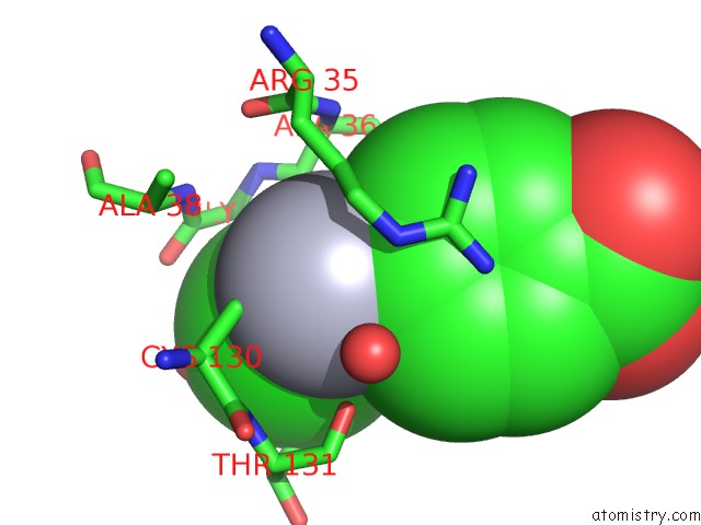
Mono view
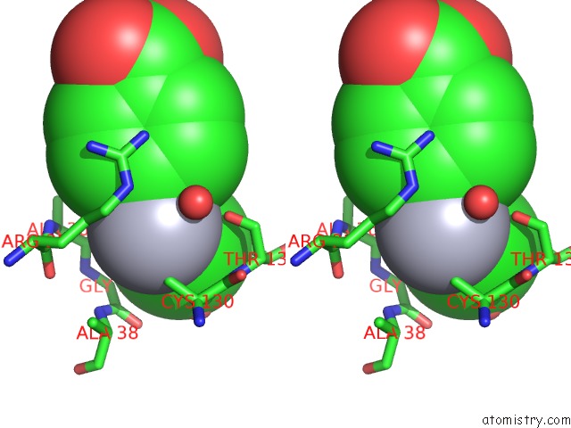
Stereo pair view
Mono view
Stereo pair view
A full contact list of Mercury with other atoms in the Hg binding
site number 1 of Crystal Structure of Struthiocalcin-1, A Different Crystal Form. within 5.0Å range:
|
Reference:
R.R.Ruiz-Arellano,
F.J.Medrano,
A.Moreno,
A.Romero.
Crystal Structure of Struthiocalcin-1, An Intramineral Protein From Struthio Camelus Eggshell, in Two Different Crystal Forms. Acta Crystallogr.,Sect.D V. 71 809 2015.
ISSN: ISSN 0907-4449
DOI: 10.1107/S139900471500125X
Page generated: Sun Aug 11 05:32:42 2024
ISSN: ISSN 0907-4449
DOI: 10.1107/S139900471500125X
Last articles
Zn in 9MJ5Zn in 9HNW
Zn in 9G0L
Zn in 9FNE
Zn in 9DZN
Zn in 9E0I
Zn in 9D32
Zn in 9DAK
Zn in 8ZXC
Zn in 8ZUF