Mercury »
PDB 4q81-5c9l »
5awg »
Mercury in PDB 5awg: Crystal Structure of Hg-Bound Sufb-Sufc-Sufd Complex From Escherichia Coli
Protein crystallography data
The structure of Crystal Structure of Hg-Bound Sufb-Sufc-Sufd Complex From Escherichia Coli, PDB code: 5awg
was solved by
K.Hirabayashi,
K.Wada,
with X-Ray Crystallography technique. A brief refinement statistics is given in the table below:
| Resolution Low / High (Å) | 43.94 / 4.28 |
| Space group | P 1 21 1 |
| Cell size a, b, c (Å), α, β, γ (°) | 119.848, 139.379, 124.414, 90.00, 113.55, 90.00 |
| R / Rfree (%) | 29.5 / 34 |
Mercury Binding Sites:
The binding sites of Mercury atom in the Crystal Structure of Hg-Bound Sufb-Sufc-Sufd Complex From Escherichia Coli
(pdb code 5awg). This binding sites where shown within
5.0 Angstroms radius around Mercury atom.
In total 4 binding sites of Mercury where determined in the Crystal Structure of Hg-Bound Sufb-Sufc-Sufd Complex From Escherichia Coli, PDB code: 5awg:
Jump to Mercury binding site number: 1; 2; 3; 4;
In total 4 binding sites of Mercury where determined in the Crystal Structure of Hg-Bound Sufb-Sufc-Sufd Complex From Escherichia Coli, PDB code: 5awg:
Jump to Mercury binding site number: 1; 2; 3; 4;
Mercury binding site 1 out of 4 in 5awg
Go back to
Mercury binding site 1 out
of 4 in the Crystal Structure of Hg-Bound Sufb-Sufc-Sufd Complex From Escherichia Coli

Mono view
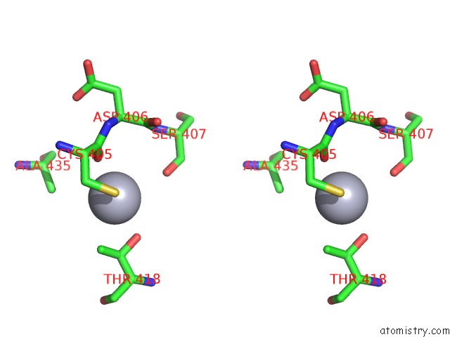
Stereo pair view
Mono view
Stereo pair view
A full contact list of Mercury with other atoms in the Hg binding
site number 1 of Crystal Structure of Hg-Bound Sufb-Sufc-Sufd Complex From Escherichia Coli within 5.0Å range:
|
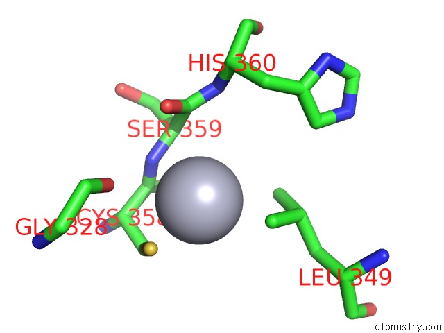
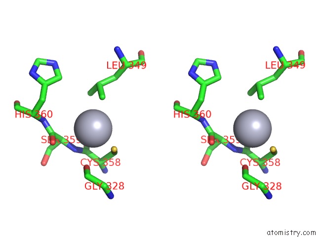
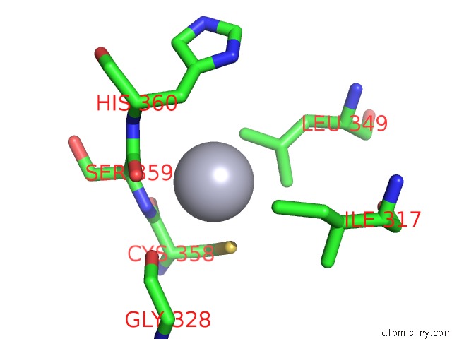
Mercury binding site 2 out of 4 in 5awg
Go back to
Mercury binding site 2 out
of 4 in the Crystal Structure of Hg-Bound Sufb-Sufc-Sufd Complex From Escherichia Coli
Mono view
Stereo pair view
Mono view
Stereo pair view
A full contact list of Mercury with other atoms in the Hg binding
site number 2 of Crystal Structure of Hg-Bound Sufb-Sufc-Sufd Complex From Escherichia Coli within 5.0Å range:
|
Mercury binding site 3 out of 4 in 5awg
Go back to
Mercury binding site 3 out
of 4 in the Crystal Structure of Hg-Bound Sufb-Sufc-Sufd Complex From Escherichia Coli

Mono view

Stereo pair view
Mono view
Stereo pair view
A full contact list of Mercury with other atoms in the Hg binding
site number 3 of Crystal Structure of Hg-Bound Sufb-Sufc-Sufd Complex From Escherichia Coli within 5.0Å range:
|
Mercury binding site 4 out of 4 in 5awg
Go back to
Mercury binding site 4 out
of 4 in the Crystal Structure of Hg-Bound Sufb-Sufc-Sufd Complex From Escherichia Coli
Mono view

Stereo pair view
Mono view
Stereo pair view
A full contact list of Mercury with other atoms in the Hg binding
site number 4 of Crystal Structure of Hg-Bound Sufb-Sufc-Sufd Complex From Escherichia Coli within 5.0Å range:
|
Reference:
K.Hirabayashi,
E.Yuda,
N.Tanaka,
S.Katayama,
K.Iwasaki,
T.Matsumoto,
G.Kurisu,
F.W.Outten,
K.Fukuyama,
Y.Takahashi,
K.Wada.
Functional Dynamics Revealed By the Structure of the Sufbcd Complex, A Novel Atp-Binding Cassette (Abc) Protein That Serves As A Scaffold For Iron-Sulfur Cluster Biogenesis J.Biol.Chem. V. 290 29717 2015.
ISSN: ESSN 1083-351X
PubMed: 26472926
DOI: 10.1074/JBC.M115.680934
Page generated: Sun Aug 11 05:39:23 2024
ISSN: ESSN 1083-351X
PubMed: 26472926
DOI: 10.1074/JBC.M115.680934
Last articles
Fe in 2YXOFe in 2YRS
Fe in 2YXC
Fe in 2YNM
Fe in 2YVJ
Fe in 2YP1
Fe in 2YU2
Fe in 2YU1
Fe in 2YQB
Fe in 2YOO