Mercury »
PDB 4q81-5c9l »
5c9l »
Mercury in PDB 5c9l: Crystal Structure of Native Pll Lectin From Photorhabdus Luminescens at 1.65 A Resolution
Protein crystallography data
The structure of Crystal Structure of Native Pll Lectin From Photorhabdus Luminescens at 1.65 A Resolution, PDB code: 5c9l
was solved by
A.Kumar,
P.Sykorova,
G.Demo,
P.Dobes,
P.Hyrsl,
M.Wimmerova,
with X-Ray Crystallography technique. A brief refinement statistics is given in the table below:
| Resolution Low / High (Å) | 39.15 / 1.65 |
| Space group | I 2 2 2 |
| Cell size a, b, c (Å), α, β, γ (°) | 71.120, 87.677, 157.691, 90.00, 90.00, 90.00 |
| R / Rfree (%) | 14.1 / 15.9 |
Other elements in 5c9l:
The structure of Crystal Structure of Native Pll Lectin From Photorhabdus Luminescens at 1.65 A Resolution also contains other interesting chemical elements:
| Chlorine | (Cl) | 1 atom |
| Calcium | (Ca) | 2 atoms |
Mercury Binding Sites:
The binding sites of Mercury atom in the Crystal Structure of Native Pll Lectin From Photorhabdus Luminescens at 1.65 A Resolution
(pdb code 5c9l). This binding sites where shown within
5.0 Angstroms radius around Mercury atom.
In total 2 binding sites of Mercury where determined in the Crystal Structure of Native Pll Lectin From Photorhabdus Luminescens at 1.65 A Resolution, PDB code: 5c9l:
Jump to Mercury binding site number: 1; 2;
In total 2 binding sites of Mercury where determined in the Crystal Structure of Native Pll Lectin From Photorhabdus Luminescens at 1.65 A Resolution, PDB code: 5c9l:
Jump to Mercury binding site number: 1; 2;
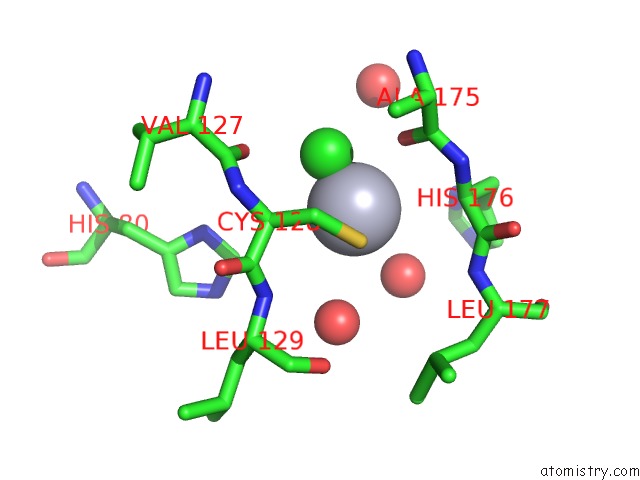
Mercury binding site 1 out of 2 in 5c9l
Go back to
Mercury binding site 1 out
of 2 in the Crystal Structure of Native Pll Lectin From Photorhabdus Luminescens at 1.65 A Resolution
Mono view
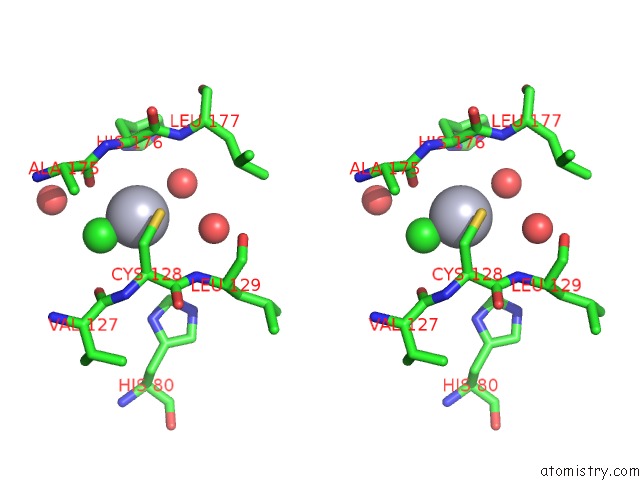
Stereo pair view
Mono view
Stereo pair view
A full contact list of Mercury with other atoms in the Hg binding
site number 1 of Crystal Structure of Native Pll Lectin From Photorhabdus Luminescens at 1.65 A Resolution within 5.0Å range:
|
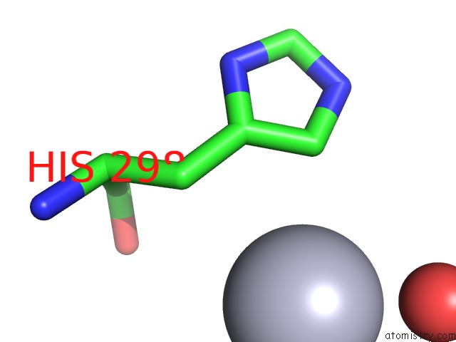
Mercury binding site 2 out of 2 in 5c9l
Go back to
Mercury binding site 2 out
of 2 in the Crystal Structure of Native Pll Lectin From Photorhabdus Luminescens at 1.65 A Resolution
Mono view
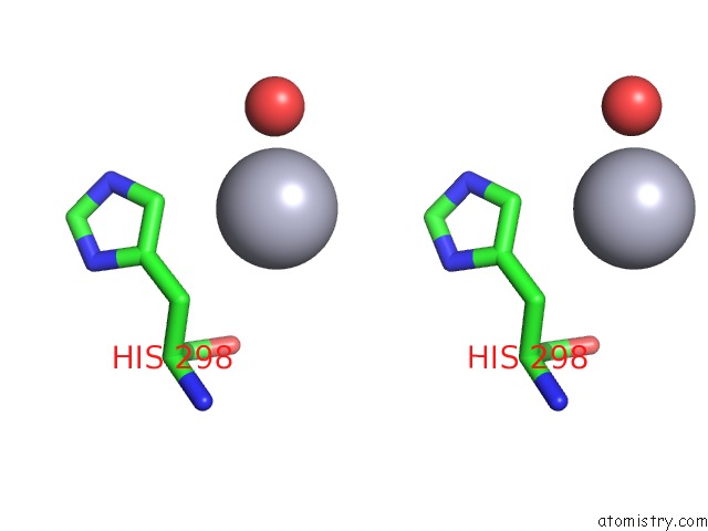
Stereo pair view
Mono view
Stereo pair view
A full contact list of Mercury with other atoms in the Hg binding
site number 2 of Crystal Structure of Native Pll Lectin From Photorhabdus Luminescens at 1.65 A Resolution within 5.0Å range:
|
Reference:
A.Kumar,
P.Sykorova,
G.Demo,
P.Dobes,
P.Hyrsl,
M.Wimmerova.
A Novel Fucose-Binding Lectin From Photorhabdus Luminescens (Pll) with An Unusual Heptabladed Beta-Propeller Tetrameric Structure. J.Biol.Chem. V. 291 25032 2016.
ISSN: ESSN 1083-351X
PubMed: 27758853
DOI: 10.1074/JBC.M115.693473
Page generated: Sun Aug 11 05:41:40 2024
ISSN: ESSN 1083-351X
PubMed: 27758853
DOI: 10.1074/JBC.M115.693473
Last articles
Zn in 9MJ5Zn in 9HNW
Zn in 9G0L
Zn in 9FNE
Zn in 9DZN
Zn in 9E0I
Zn in 9D32
Zn in 9DAK
Zn in 8ZXC
Zn in 8ZUF