Mercury »
PDB 5ca2-5l9w »
5jkn »
Mercury in PDB 5jkn: Crystal Structure of Deubiquitinase Mindy-1
Protein crystallography data
The structure of Crystal Structure of Deubiquitinase Mindy-1, PDB code: 5jkn
was solved by
S.A.Abdul Rehman,
Y.Kulathu,
with X-Ray Crystallography technique. A brief refinement statistics is given in the table below:
| Resolution Low / High (Å) | 99.67 / 3.00 |
| Space group | P 41 2 2 |
| Cell size a, b, c (Å), α, β, γ (°) | 99.671, 99.671, 165.123, 90.00, 90.00, 90.00 |
| R / Rfree (%) | 19.7 / 24.2 |
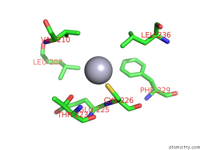
Mercury Binding Sites:
The binding sites of Mercury atom in the Crystal Structure of Deubiquitinase Mindy-1
(pdb code 5jkn). This binding sites where shown within
5.0 Angstroms radius around Mercury atom.
In total only one binding site of Mercury was determined in the Crystal Structure of Deubiquitinase Mindy-1, PDB code: 5jkn:
In total only one binding site of Mercury was determined in the Crystal Structure of Deubiquitinase Mindy-1, PDB code: 5jkn:
Mercury binding site 1 out of 1 in 5jkn
Go back to
Mercury binding site 1 out
of 1 in the Crystal Structure of Deubiquitinase Mindy-1
Mono view
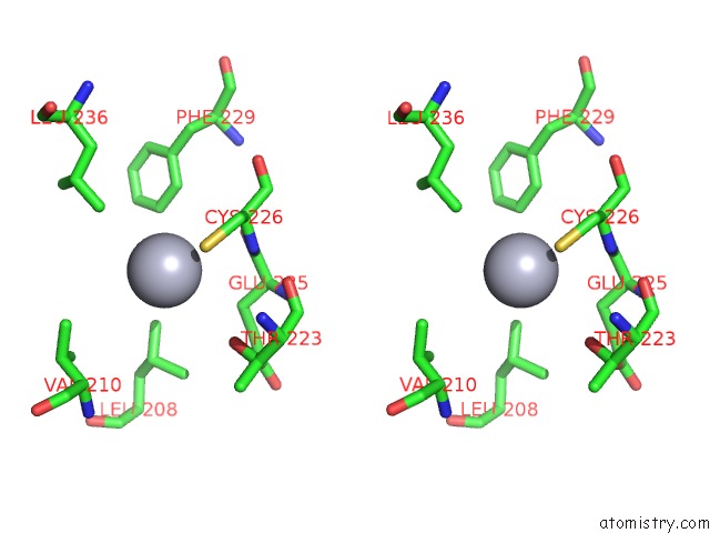
Stereo pair view
Mono view
Stereo pair view
A full contact list of Mercury with other atoms in the Hg binding
site number 1 of Crystal Structure of Deubiquitinase Mindy-1 within 5.0Å range:
|
Reference:
S.A.Abdul Rehman,
Y.A.Kristariyanto,
S.Y.Choi,
P.J.Nkosi,
S.Weidlich,
K.Labib,
K.Hofmann,
Y.Kulathu.
Mindy-1 Is A Member of An Evolutionarily Conserved and Structurally Distinct New Family of Deubiquitinating Enzymes. Mol.Cell V. 63 146 2016.
ISSN: ISSN 1097-2765
PubMed: 27292798
DOI: 10.1016/J.MOLCEL.2016.05.009
Page generated: Sun Aug 11 06:26:20 2024
ISSN: ISSN 1097-2765
PubMed: 27292798
DOI: 10.1016/J.MOLCEL.2016.05.009
Last articles
Cl in 5R8UCl in 5R7X
Cl in 5R7Z
Cl in 5R5U
Cl in 5R5Q
Cl in 5R7Y
Cl in 5QTP
Cl in 5R5N
Cl in 5R5H
Cl in 5QU0