Mercury »
PDB 5lf8-6bvg »
5mjo »
Mercury in PDB 5mjo: Structure of PSB29 at 1.55A
Protein crystallography data
The structure of Structure of PSB29 at 1.55A, PDB code: 5mjo
was solved by
J.W.Murray,
A.Kozlo,
with X-Ray Crystallography technique. A brief refinement statistics is given in the table below:
| Resolution Low / High (Å) | 46.16 / 1.55 |
| Space group | P 1 21 1 |
| Cell size a, b, c (Å), α, β, γ (°) | 31.240, 56.730, 47.730, 90.00, 104.72, 90.00 |
| R / Rfree (%) | 13.3 / 19.2 |
Other elements in 5mjo:
The structure of Structure of PSB29 at 1.55A also contains other interesting chemical elements:
| Iodine | (I) | 2 atoms |
Mercury Binding Sites:
The binding sites of Mercury atom in the Structure of PSB29 at 1.55A
(pdb code 5mjo). This binding sites where shown within
5.0 Angstroms radius around Mercury atom.
In total only one binding site of Mercury was determined in the Structure of PSB29 at 1.55A, PDB code: 5mjo:
In total only one binding site of Mercury was determined in the Structure of PSB29 at 1.55A, PDB code: 5mjo:
Mercury binding site 1 out of 1 in 5mjo
Go back to
Mercury binding site 1 out
of 1 in the Structure of PSB29 at 1.55A
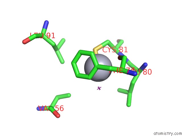
Mono view
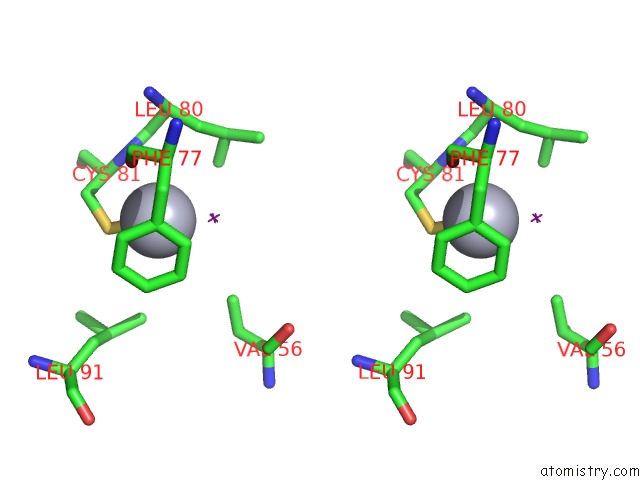
Stereo pair view
Mono view
Stereo pair view
A full contact list of Mercury with other atoms in the Hg binding
site number 1 of Structure of PSB29 at 1.55A within 5.0Å range:
|
Reference:
M.Bec Kova,
J.Yu,
V.Krynicka,
A.Kozlo,
S.Shao,
P.Konik,
J.Komenda,
J.W.Murray,
P.J.Nixon.
Structure of PSB29/THF1 and Its Association with the Ftsh Protease Complex Involved in Photosystem II Repair in Cyanobacteria. Philos. Trans. R. Soc. V. 372 2017LOND., B, Biol. Sci..
ISSN: ESSN 1471-2970
PubMed: 28808107
DOI: 10.1098/RSTB.2016.0394
Page generated: Sun Aug 11 06:50:31 2024
ISSN: ESSN 1471-2970
PubMed: 28808107
DOI: 10.1098/RSTB.2016.0394
Last articles
Zn in 9MJ5Zn in 9HNW
Zn in 9G0L
Zn in 9FNE
Zn in 9DZN
Zn in 9E0I
Zn in 9D32
Zn in 9DAK
Zn in 8ZXC
Zn in 8ZUF