Mercury »
PDB 5lf8-6bvg »
5n83 »
Mercury in PDB 5n83: Structure of the Distal Domain of Mouse Adenovirus 2 Fibre, Methylmercury Chloride Derivative
Protein crystallography data
The structure of Structure of the Distal Domain of Mouse Adenovirus 2 Fibre, Methylmercury Chloride Derivative, PDB code: 5n83
was solved by
A.K.Singh,
M.J.Van Raaij,
with X-Ray Crystallography technique. A brief refinement statistics is given in the table below:
| Resolution Low / High (Å) | 82.14 / 2.76 |
| Space group | C 2 2 21 |
| Cell size a, b, c (Å), α, β, γ (°) | 92.116, 164.285, 96.432, 90.00, 90.00, 90.00 |
| R / Rfree (%) | 20.3 / 24.8 |
Mercury Binding Sites:
The binding sites of Mercury atom in the Structure of the Distal Domain of Mouse Adenovirus 2 Fibre, Methylmercury Chloride Derivative
(pdb code 5n83). This binding sites where shown within
5.0 Angstroms radius around Mercury atom.
In total 5 binding sites of Mercury where determined in the Structure of the Distal Domain of Mouse Adenovirus 2 Fibre, Methylmercury Chloride Derivative, PDB code: 5n83:
Jump to Mercury binding site number: 1; 2; 3; 4; 5;
In total 5 binding sites of Mercury where determined in the Structure of the Distal Domain of Mouse Adenovirus 2 Fibre, Methylmercury Chloride Derivative, PDB code: 5n83:
Jump to Mercury binding site number: 1; 2; 3; 4; 5;
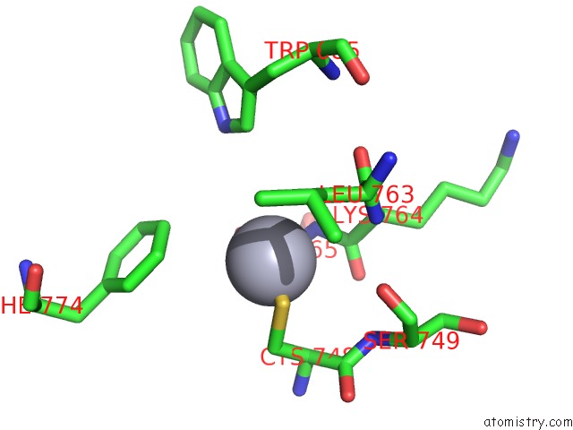
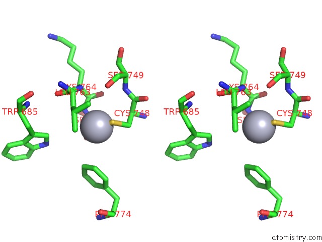
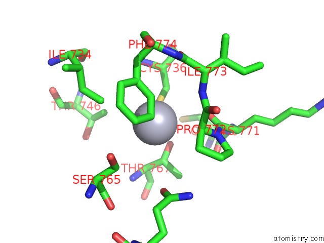
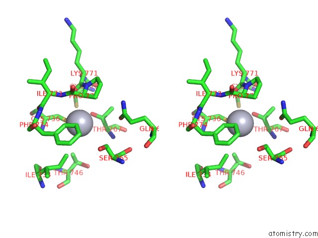
Mercury binding site 1 out of 5 in 5n83
Go back to
Mercury binding site 1 out
of 5 in the Structure of the Distal Domain of Mouse Adenovirus 2 Fibre, Methylmercury Chloride Derivative
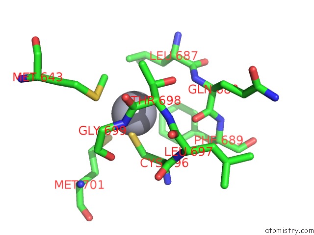
Mono view
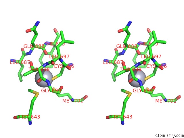
Stereo pair view
Mono view
Stereo pair view
A full contact list of Mercury with other atoms in the Hg binding
site number 1 of Structure of the Distal Domain of Mouse Adenovirus 2 Fibre, Methylmercury Chloride Derivative within 5.0Å range:
|
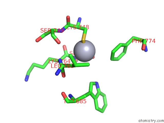
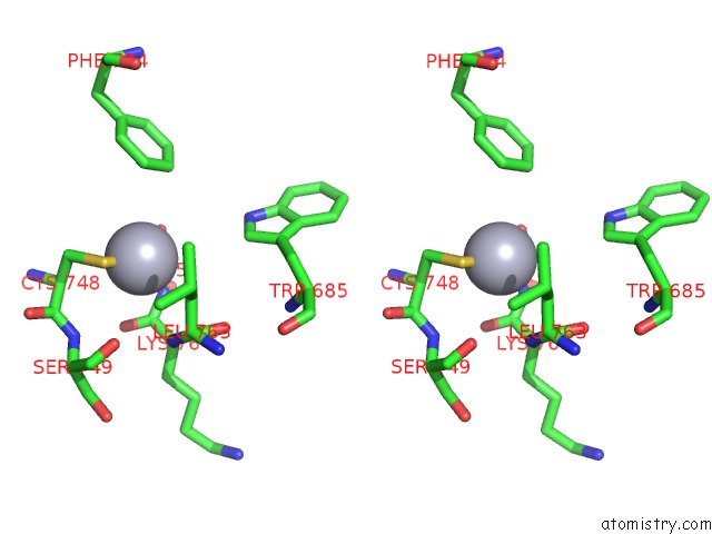
Mercury binding site 2 out of 5 in 5n83
Go back to
Mercury binding site 2 out
of 5 in the Structure of the Distal Domain of Mouse Adenovirus 2 Fibre, Methylmercury Chloride Derivative
Mono view
Stereo pair view
Mono view
Stereo pair view
A full contact list of Mercury with other atoms in the Hg binding
site number 2 of Structure of the Distal Domain of Mouse Adenovirus 2 Fibre, Methylmercury Chloride Derivative within 5.0Å range:
|
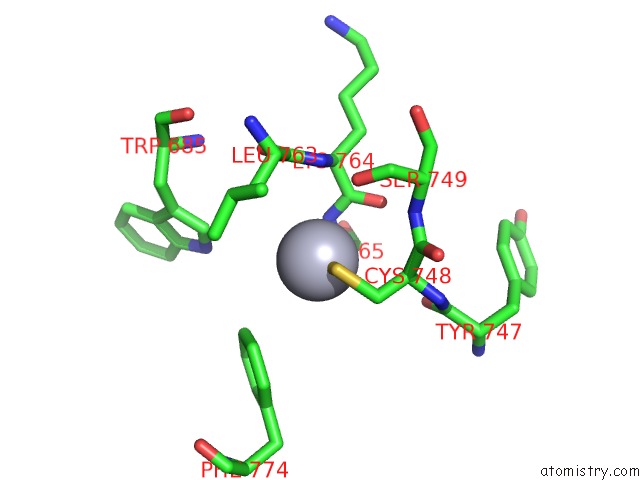
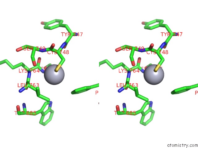
Mercury binding site 3 out of 5 in 5n83
Go back to
Mercury binding site 3 out
of 5 in the Structure of the Distal Domain of Mouse Adenovirus 2 Fibre, Methylmercury Chloride Derivative
Mono view
Stereo pair view
Mono view
Stereo pair view
A full contact list of Mercury with other atoms in the Hg binding
site number 3 of Structure of the Distal Domain of Mouse Adenovirus 2 Fibre, Methylmercury Chloride Derivative within 5.0Å range:
|
Mercury binding site 4 out of 5 in 5n83
Go back to
Mercury binding site 4 out
of 5 in the Structure of the Distal Domain of Mouse Adenovirus 2 Fibre, Methylmercury Chloride Derivative
Mono view
Stereo pair view
Mono view
Stereo pair view
A full contact list of Mercury with other atoms in the Hg binding
site number 4 of Structure of the Distal Domain of Mouse Adenovirus 2 Fibre, Methylmercury Chloride Derivative within 5.0Å range:
|
Mercury binding site 5 out of 5 in 5n83
Go back to
Mercury binding site 5 out
of 5 in the Structure of the Distal Domain of Mouse Adenovirus 2 Fibre, Methylmercury Chloride Derivative
Mono view
Stereo pair view
Mono view
Stereo pair view
A full contact list of Mercury with other atoms in the Hg binding
site number 5 of Structure of the Distal Domain of Mouse Adenovirus 2 Fibre, Methylmercury Chloride Derivative within 5.0Å range:
|
Reference:
A.K.Singh,
T.H.Nguyen,
M.Z.Vidovszky,
B.Harrach,
M.Benko,
A.Kirwan,
L.Joshi,
M.Kilcoyne,
M.A.Berbis,
F.J.Canada,
J.Jimenez-Barbero,
M.Menendez,
S.S.Wilson,
B.A.Bromme,
J.G.Smith,
M.J.Van Raaij.
Structure and N-Acetylglucosamine Binding of the Distal Domain of Mouse Adenovirus 2 Fibre. J. Gen. Virol. V. 99 1494 2018.
ISSN: ESSN 1465-2099
PubMed: 30277856
DOI: 10.1099/JGV.0.001145
Page generated: Sun Aug 11 06:50:31 2024
ISSN: ESSN 1465-2099
PubMed: 30277856
DOI: 10.1099/JGV.0.001145
Last articles
Cl in 5RAOCl in 5RAN
Cl in 5RAJ
Cl in 5RAM
Cl in 5RAL
Cl in 5RAK
Cl in 5RAF
Cl in 5RAG
Cl in 5RAH
Cl in 5RAI