Mercury »
PDB 5lf8-6bvg »
5oon »
Mercury in PDB 5oon: Structure of Undecaprenyl-Pyrophosphate Phosphatase, Baca
Enzymatic activity of Structure of Undecaprenyl-Pyrophosphate Phosphatase, Baca
All present enzymatic activity of Structure of Undecaprenyl-Pyrophosphate Phosphatase, Baca:
3.6.1.27;
3.6.1.27;
Protein crystallography data
The structure of Structure of Undecaprenyl-Pyrophosphate Phosphatase, Baca, PDB code: 5oon
was solved by
C.-Y.Huang,
V.Olieric,
R.Warshamanage,
M.Wang,
N.Howe,
M.E.I.Ghachi,
D.Weichert,
F.Kerff,
P.Stansfeld,
T.Touze,
M.Caffrey,
with X-Ray Crystallography technique. A brief refinement statistics is given in the table below:
| Resolution Low / High (Å) | 44.46 / 2.60 |
| Space group | C 2 2 2 |
| Cell size a, b, c (Å), α, β, γ (°) | 113.260, 145.000, 40.490, 90.00, 90.00, 90.00 |
| R / Rfree (%) | 20.6 / 24.4 |
Mercury Binding Sites:
The binding sites of Mercury atom in the Structure of Undecaprenyl-Pyrophosphate Phosphatase, Baca
(pdb code 5oon). This binding sites where shown within
5.0 Angstroms radius around Mercury atom.
In total 2 binding sites of Mercury where determined in the Structure of Undecaprenyl-Pyrophosphate Phosphatase, Baca, PDB code: 5oon:
Jump to Mercury binding site number: 1; 2;
In total 2 binding sites of Mercury where determined in the Structure of Undecaprenyl-Pyrophosphate Phosphatase, Baca, PDB code: 5oon:
Jump to Mercury binding site number: 1; 2;
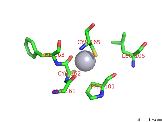
Mercury binding site 1 out of 2 in 5oon
Go back to
Mercury binding site 1 out
of 2 in the Structure of Undecaprenyl-Pyrophosphate Phosphatase, Baca
Mono view
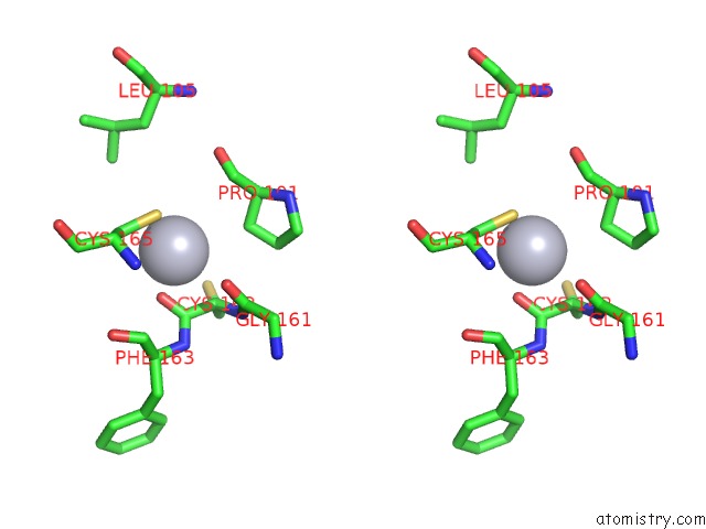
Stereo pair view
Mono view
Stereo pair view
A full contact list of Mercury with other atoms in the Hg binding
site number 1 of Structure of Undecaprenyl-Pyrophosphate Phosphatase, Baca within 5.0Å range:
|
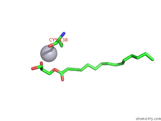
Mercury binding site 2 out of 2 in 5oon
Go back to
Mercury binding site 2 out
of 2 in the Structure of Undecaprenyl-Pyrophosphate Phosphatase, Baca
Mono view
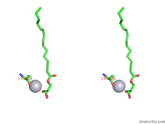
Stereo pair view
Mono view
Stereo pair view
A full contact list of Mercury with other atoms in the Hg binding
site number 2 of Structure of Undecaprenyl-Pyrophosphate Phosphatase, Baca within 5.0Å range:
|
Reference:
M.El Ghachi,
N.Howe,
C.Y.Huang,
V.Olieric,
R.Warshamanage,
T.Touze,
D.Weichert,
P.J.Stansfeld,
M.Wang,
F.Kerff,
M.Caffrey.
Crystal Structure of Undecaprenyl-Pyrophosphate Phosphatase and Its Role in Peptidoglycan Biosynthesis. Nat Commun V. 9 1078 2018.
ISSN: ESSN 2041-1723
PubMed: 29540682
DOI: 10.1038/S41467-018-03477-5
Page generated: Sun Aug 11 06:50:31 2024
ISSN: ESSN 2041-1723
PubMed: 29540682
DOI: 10.1038/S41467-018-03477-5
Last articles
Cl in 3O6XCl in 3O9L
Cl in 3OA0
Cl in 3O9Z
Cl in 3O8M
Cl in 3O8U
Cl in 3O8T
Cl in 3O8P
Cl in 3O79
Cl in 3O6O