Mercury »
PDB 5lf8-6bvg »
5tiq »
Mercury in PDB 5tiq: The Structure of the Major Capsid Protein of Pbcv-1
Protein crystallography data
The structure of The Structure of the Major Capsid Protein of Pbcv-1, PDB code: 5tiq
was solved by
T.Klose,
C.De Castro,
I.Speciale,
A.Molinaro,
J.L.Van Etten,
M.G.Rossmann,
with X-Ray Crystallography technique. A brief refinement statistics is given in the table below:
| Resolution Low / High (Å) | 43.31 / 2.54 |
| Space group | P 41 3 2 |
| Cell size a, b, c (Å), α, β, γ (°) | 188.763, 188.763, 188.763, 90.00, 90.00, 90.00 |
| R / Rfree (%) | 17.9 / 22.3 |
Mercury Binding Sites:
The binding sites of Mercury atom in the The Structure of the Major Capsid Protein of Pbcv-1
(pdb code 5tiq). This binding sites where shown within
5.0 Angstroms radius around Mercury atom.
In total 4 binding sites of Mercury where determined in the The Structure of the Major Capsid Protein of Pbcv-1, PDB code: 5tiq:
Jump to Mercury binding site number: 1; 2; 3; 4;
In total 4 binding sites of Mercury where determined in the The Structure of the Major Capsid Protein of Pbcv-1, PDB code: 5tiq:
Jump to Mercury binding site number: 1; 2; 3; 4;
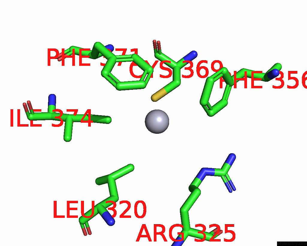
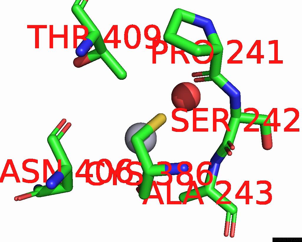
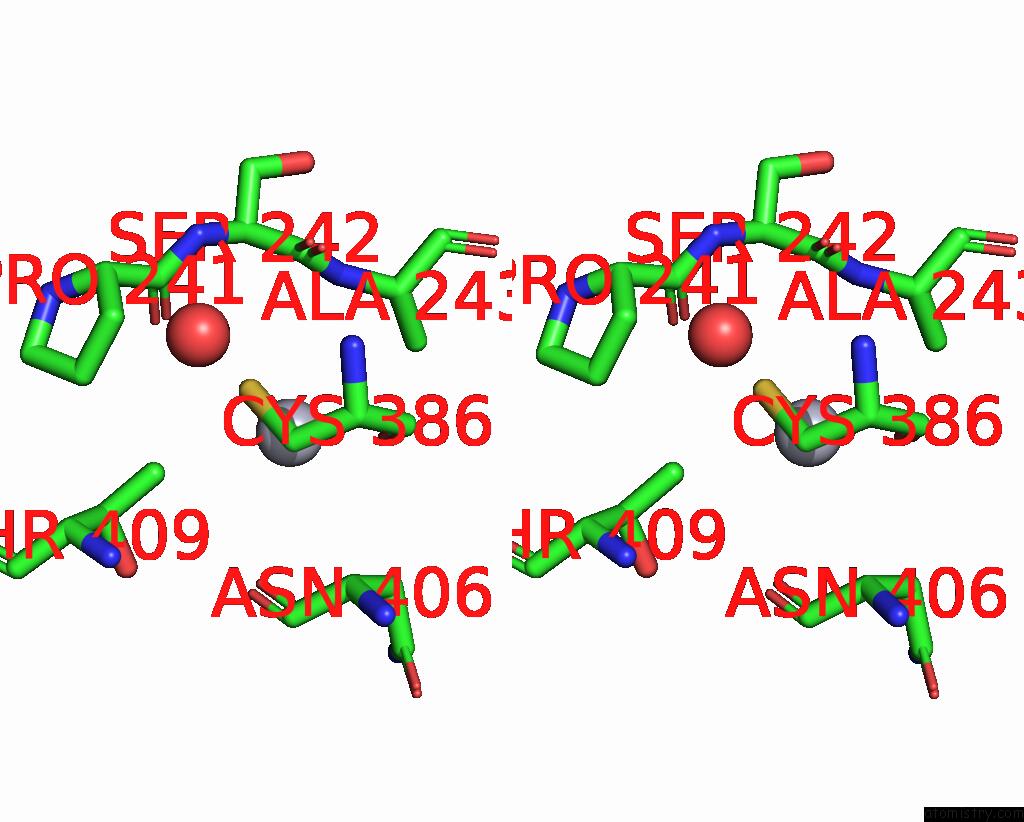
Mercury binding site 1 out of 4 in 5tiq
Go back to
Mercury binding site 1 out
of 4 in the The Structure of the Major Capsid Protein of Pbcv-1
Mono view
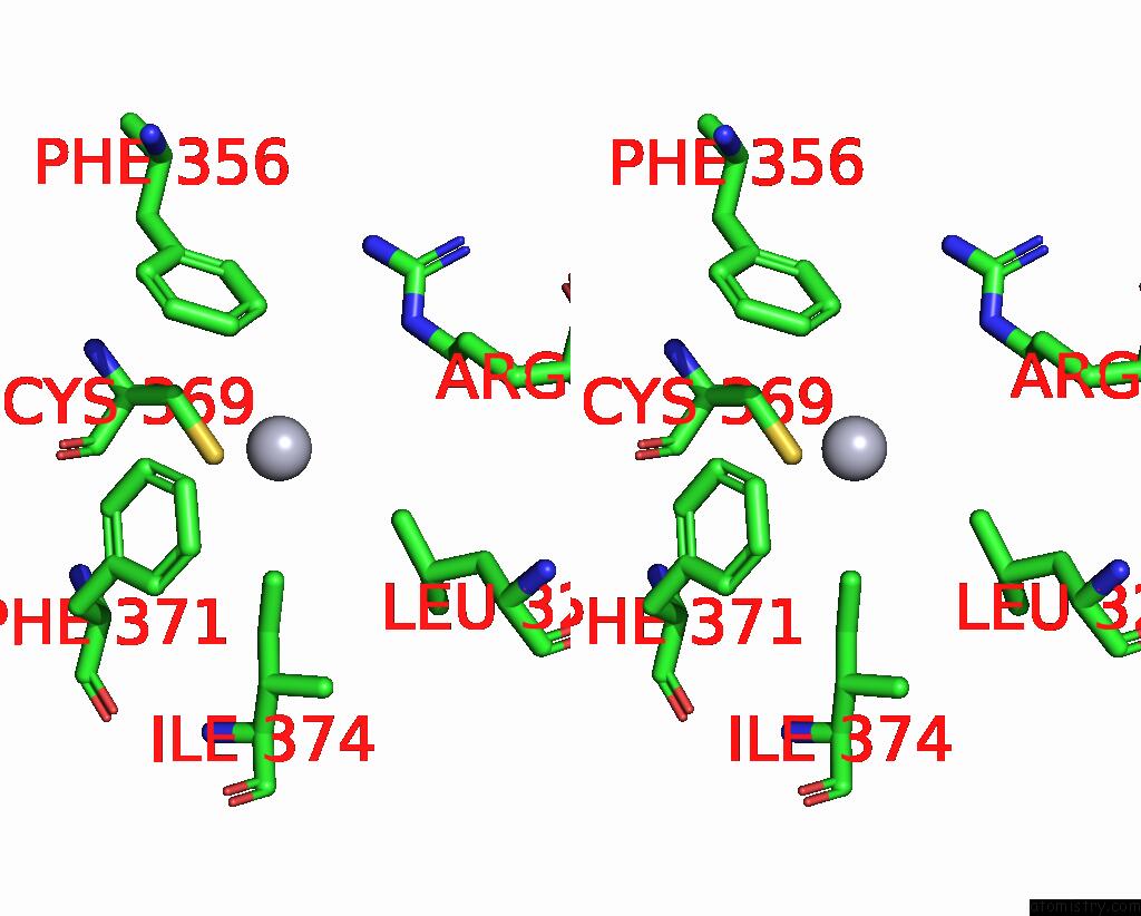
Stereo pair view
Mono view
Stereo pair view
A full contact list of Mercury with other atoms in the Hg binding
site number 1 of The Structure of the Major Capsid Protein of Pbcv-1 within 5.0Å range:
|
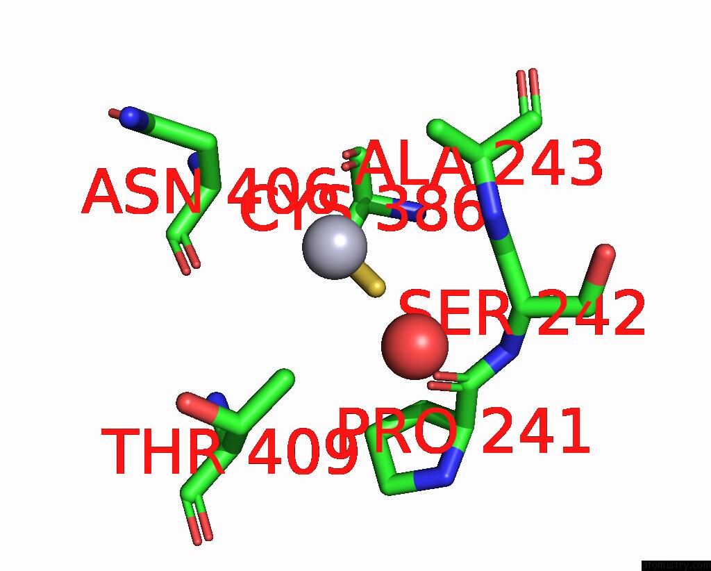
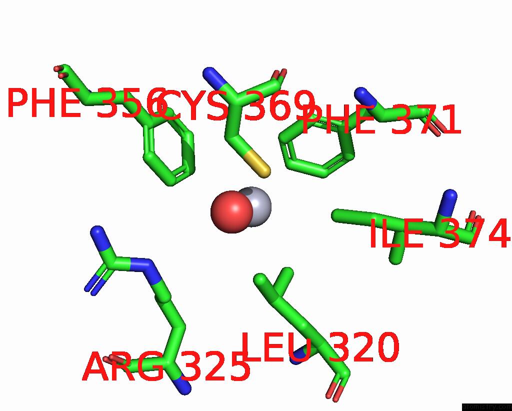
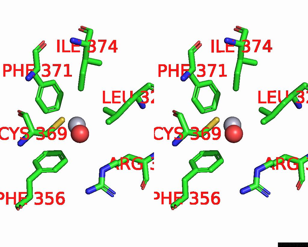
Mercury binding site 2 out of 4 in 5tiq
Go back to
Mercury binding site 2 out
of 4 in the The Structure of the Major Capsid Protein of Pbcv-1
Mono view
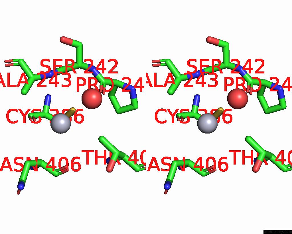
Stereo pair view
Mono view
Stereo pair view
A full contact list of Mercury with other atoms in the Hg binding
site number 2 of The Structure of the Major Capsid Protein of Pbcv-1 within 5.0Å range:
|
Mercury binding site 3 out of 4 in 5tiq
Go back to
Mercury binding site 3 out
of 4 in the The Structure of the Major Capsid Protein of Pbcv-1
Mono view
Stereo pair view
Mono view
Stereo pair view
A full contact list of Mercury with other atoms in the Hg binding
site number 3 of The Structure of the Major Capsid Protein of Pbcv-1 within 5.0Å range:
|
Mercury binding site 4 out of 4 in 5tiq
Go back to
Mercury binding site 4 out
of 4 in the The Structure of the Major Capsid Protein of Pbcv-1
Mono view
Stereo pair view
Mono view
Stereo pair view
A full contact list of Mercury with other atoms in the Hg binding
site number 4 of The Structure of the Major Capsid Protein of Pbcv-1 within 5.0Å range:
|
Reference:
C.De Castro,
T.Klose,
I.Speciale,
R.Lanzetta,
A.Molinaro,
J.L.Van Etten,
M.G.Rossmann.
Structure of the Chlorovirus Pbcv-1 Major Capsid Glycoprotein Determined By Combining Crystallographic and Carbohydrate Molecular Modeling Approaches. Proc. Natl. Acad. Sci. V. 115 E44 2018U.S.A..
ISSN: ESSN 1091-6490
PubMed: 29255015
DOI: 10.1073/PNAS.1613432115
Page generated: Sun Aug 11 06:55:08 2024
ISSN: ESSN 1091-6490
PubMed: 29255015
DOI: 10.1073/PNAS.1613432115
Last articles
Zn in 9MJ5Zn in 9HNW
Zn in 9G0L
Zn in 9FNE
Zn in 9DZN
Zn in 9E0I
Zn in 9D32
Zn in 9DAK
Zn in 8ZXC
Zn in 8ZUF