Mercury »
PDB 5lf8-6bvg »
5wsr »
Mercury in PDB 5wsr: Crystal Structure of T-Hg-T Pair Containing Dna Duplex
Protein crystallography data
The structure of Crystal Structure of T-Hg-T Pair Containing Dna Duplex, PDB code: 5wsr
was solved by
J.H.Gan,
H.H.Liu,
with X-Ray Crystallography technique. A brief refinement statistics is given in the table below:
| Resolution Low / High (Å) | 21.57 / 1.50 |
| Space group | P 43 21 2 |
| Cell size a, b, c (Å), α, β, γ (°) | 42.524, 42.524, 25.027, 90.00, 90.00, 90.00 |
| R / Rfree (%) | 16.8 / 18.8 |
Mercury Binding Sites:
The binding sites of Mercury atom in the Crystal Structure of T-Hg-T Pair Containing Dna Duplex
(pdb code 5wsr). This binding sites where shown within
5.0 Angstroms radius around Mercury atom.
In total 2 binding sites of Mercury where determined in the Crystal Structure of T-Hg-T Pair Containing Dna Duplex, PDB code: 5wsr:
Jump to Mercury binding site number: 1; 2;
In total 2 binding sites of Mercury where determined in the Crystal Structure of T-Hg-T Pair Containing Dna Duplex, PDB code: 5wsr:
Jump to Mercury binding site number: 1; 2;
Mercury binding site 1 out of 2 in 5wsr
Go back to
Mercury binding site 1 out
of 2 in the Crystal Structure of T-Hg-T Pair Containing Dna Duplex
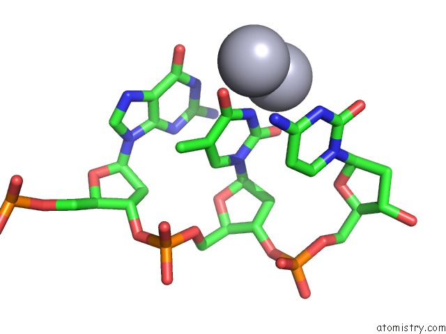
Mono view
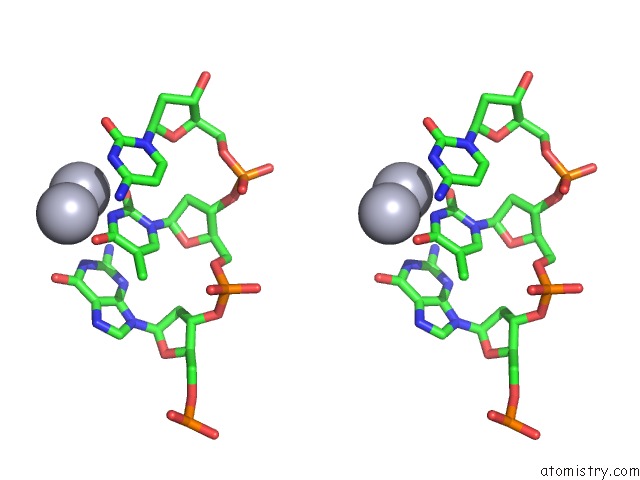
Stereo pair view
Mono view
Stereo pair view
A full contact list of Mercury with other atoms in the Hg binding
site number 1 of Crystal Structure of T-Hg-T Pair Containing Dna Duplex within 5.0Å range:
|
Mercury binding site 2 out of 2 in 5wsr
Go back to
Mercury binding site 2 out
of 2 in the Crystal Structure of T-Hg-T Pair Containing Dna Duplex
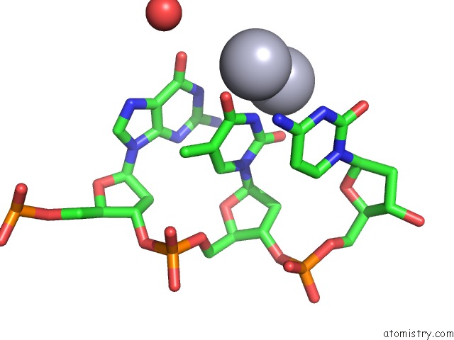
Mono view
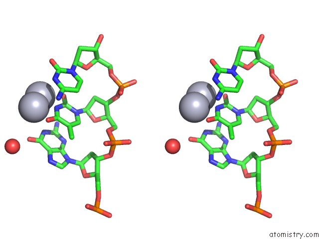
Stereo pair view
Mono view
Stereo pair view
A full contact list of Mercury with other atoms in the Hg binding
site number 2 of Crystal Structure of T-Hg-T Pair Containing Dna Duplex within 5.0Å range:
|
Reference:
H.H.Liu,
C.Cai,
P.Haruehanroengra,
Q.Q.Yao,
Y.Q.Chen,
C.Yang,
Q.Luo,
B.X.Wu,
J.X.Li,
J.B.Ma,
J.Sheng,
J.H.Gan.
Flexibility and Stabilization of Hgii-Mediated C:T and T:T Base Pairs in Dna Duplex Nucleic Acids Res. V. 45 2910 2017.
ISSN: ESSN 1362-4962
PubMed: 27998930
DOI: 10.1093/NAR/GKW1296
Page generated: Fri Aug 8 11:01:00 2025
ISSN: ESSN 1362-4962
PubMed: 27998930
DOI: 10.1093/NAR/GKW1296
Last articles
I in 4CDWI in 4BVX
I in 4BSW
I in 4AW7
I in 4BSV
I in 4B43
I in 4B9H
I in 4AS2
I in 4AS5
I in 4AX2