Mercury »
PDB 5lf8-6bvg »
5ydc »
Mercury in PDB 5ydc: Crystal Structure of Mercury Soaked C-Terminal Domain of RV1828 From Mycobacterium Tuberculosis
Protein crystallography data
The structure of Crystal Structure of Mercury Soaked C-Terminal Domain of RV1828 From Mycobacterium Tuberculosis, PDB code: 5ydc
was solved by
S.Singh,
S.Karthikeyan,
with X-Ray Crystallography technique. A brief refinement statistics is given in the table below:
| Resolution Low / High (Å) | 44.89 / 1.91 |
| Space group | P 1 21 1 |
| Cell size a, b, c (Å), α, β, γ (°) | 45.355, 31.511, 77.901, 90.00, 98.21, 90.00 |
| R / Rfree (%) | 20.6 / 26 |
Mercury Binding Sites:
The binding sites of Mercury atom in the Crystal Structure of Mercury Soaked C-Terminal Domain of RV1828 From Mycobacterium Tuberculosis
(pdb code 5ydc). This binding sites where shown within
5.0 Angstroms radius around Mercury atom.
In total 6 binding sites of Mercury where determined in the Crystal Structure of Mercury Soaked C-Terminal Domain of RV1828 From Mycobacterium Tuberculosis, PDB code: 5ydc:
Jump to Mercury binding site number: 1; 2; 3; 4; 5; 6;
In total 6 binding sites of Mercury where determined in the Crystal Structure of Mercury Soaked C-Terminal Domain of RV1828 From Mycobacterium Tuberculosis, PDB code: 5ydc:
Jump to Mercury binding site number: 1; 2; 3; 4; 5; 6;
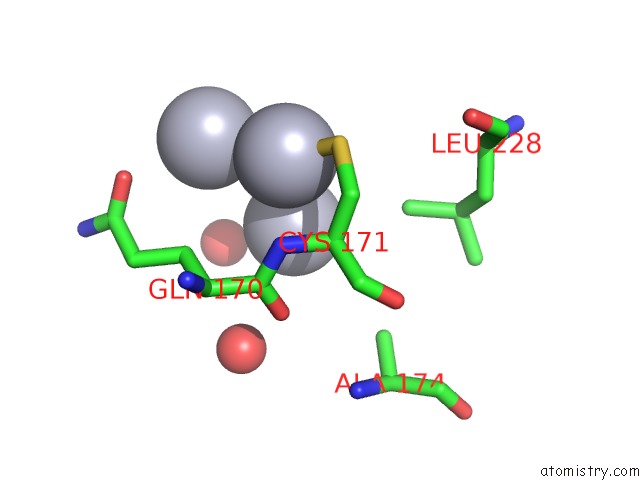
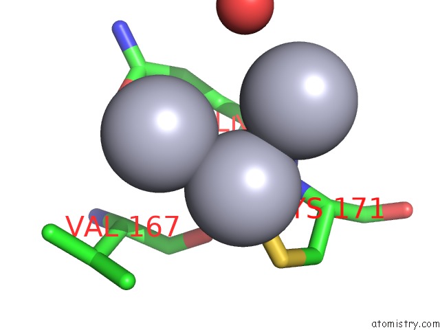
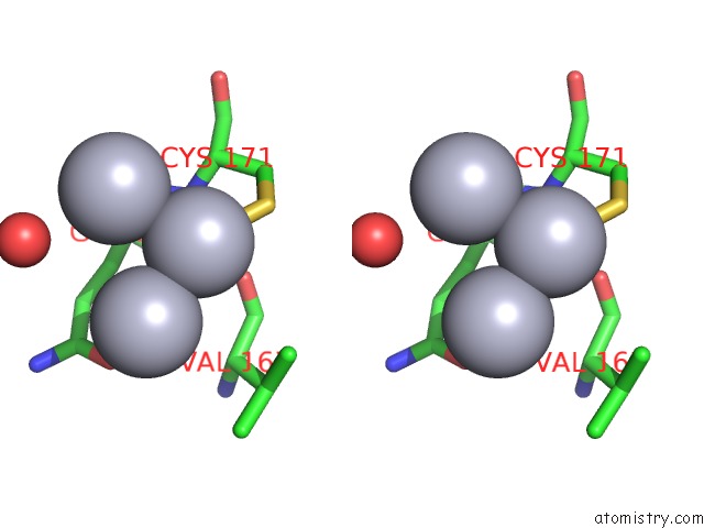
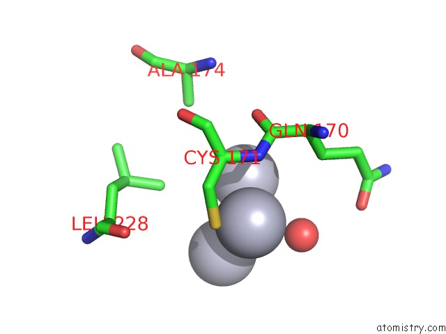
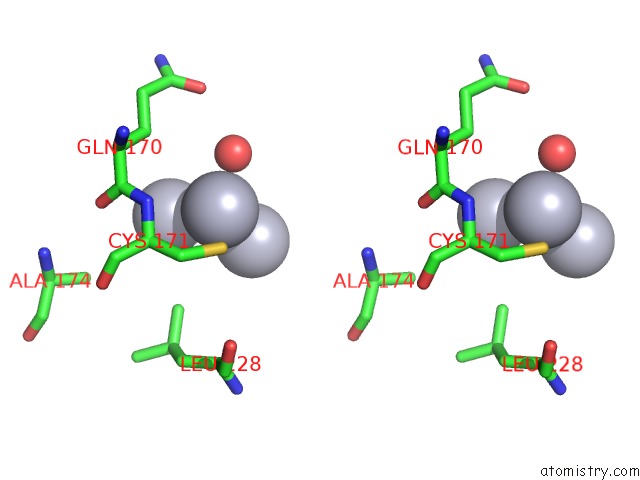
Mercury binding site 1 out of 6 in 5ydc
Go back to
Mercury binding site 1 out
of 6 in the Crystal Structure of Mercury Soaked C-Terminal Domain of RV1828 From Mycobacterium Tuberculosis
Mono view
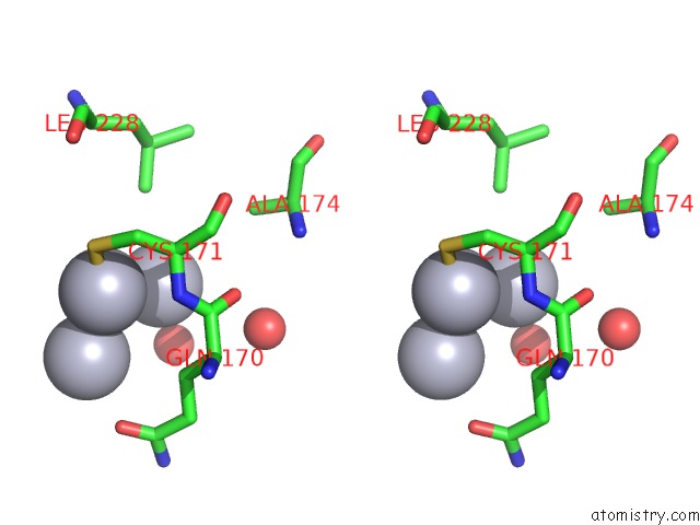
Stereo pair view
Mono view
Stereo pair view
A full contact list of Mercury with other atoms in the Hg binding
site number 1 of Crystal Structure of Mercury Soaked C-Terminal Domain of RV1828 From Mycobacterium Tuberculosis within 5.0Å range:
|
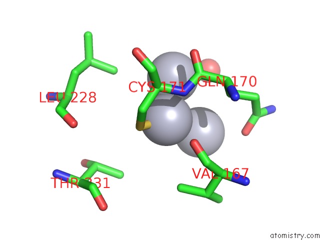
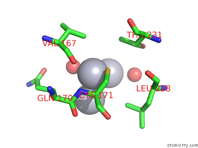
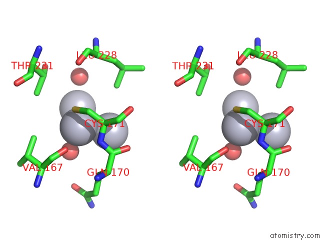
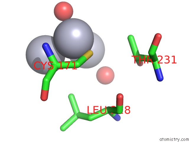
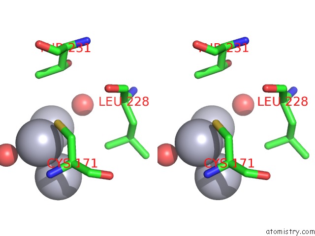
Mercury binding site 2 out of 6 in 5ydc
Go back to
Mercury binding site 2 out
of 6 in the Crystal Structure of Mercury Soaked C-Terminal Domain of RV1828 From Mycobacterium Tuberculosis
Mono view
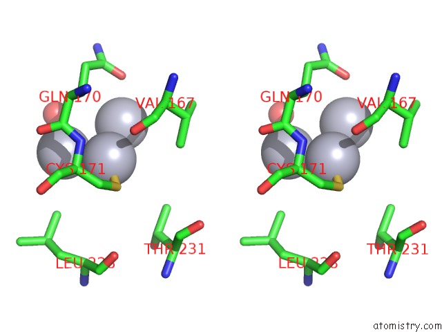
Stereo pair view
Mono view
Stereo pair view
A full contact list of Mercury with other atoms in the Hg binding
site number 2 of Crystal Structure of Mercury Soaked C-Terminal Domain of RV1828 From Mycobacterium Tuberculosis within 5.0Å range:
|
Mercury binding site 3 out of 6 in 5ydc
Go back to
Mercury binding site 3 out
of 6 in the Crystal Structure of Mercury Soaked C-Terminal Domain of RV1828 From Mycobacterium Tuberculosis
Mono view
Stereo pair view
Mono view
Stereo pair view
A full contact list of Mercury with other atoms in the Hg binding
site number 3 of Crystal Structure of Mercury Soaked C-Terminal Domain of RV1828 From Mycobacterium Tuberculosis within 5.0Å range:
|
Mercury binding site 4 out of 6 in 5ydc
Go back to
Mercury binding site 4 out
of 6 in the Crystal Structure of Mercury Soaked C-Terminal Domain of RV1828 From Mycobacterium Tuberculosis
Mono view
Stereo pair view
Mono view
Stereo pair view
A full contact list of Mercury with other atoms in the Hg binding
site number 4 of Crystal Structure of Mercury Soaked C-Terminal Domain of RV1828 From Mycobacterium Tuberculosis within 5.0Å range:
|
Mercury binding site 5 out of 6 in 5ydc
Go back to
Mercury binding site 5 out
of 6 in the Crystal Structure of Mercury Soaked C-Terminal Domain of RV1828 From Mycobacterium Tuberculosis
Mono view
Stereo pair view
Mono view
Stereo pair view
A full contact list of Mercury with other atoms in the Hg binding
site number 5 of Crystal Structure of Mercury Soaked C-Terminal Domain of RV1828 From Mycobacterium Tuberculosis within 5.0Å range:
|
Mercury binding site 6 out of 6 in 5ydc
Go back to
Mercury binding site 6 out
of 6 in the Crystal Structure of Mercury Soaked C-Terminal Domain of RV1828 From Mycobacterium Tuberculosis
Mono view
Stereo pair view
Mono view
Stereo pair view
A full contact list of Mercury with other atoms in the Hg binding
site number 6 of Crystal Structure of Mercury Soaked C-Terminal Domain of RV1828 From Mycobacterium Tuberculosis within 5.0Å range:
|
Reference:
S.Singh,
R.R.Sevalkar,
D.Sarkar,
S.Karthikeyan.
Characteristics of the Essential Pathogenicity Factor RV1828, A Merr Family Transcription Regulator From Mycobacterium Tuberculosis. Febs J. V. 285 4424 2018.
ISSN: ISSN 1742-4658
PubMed: 30306715
DOI: 10.1111/FEBS.14676
Page generated: Fri Aug 8 11:01:32 2025
ISSN: ISSN 1742-4658
PubMed: 30306715
DOI: 10.1111/FEBS.14676
Last articles
I in 4CDWI in 4BVX
I in 4BSW
I in 4AW7
I in 4BSV
I in 4B43
I in 4B9H
I in 4AS2
I in 4AS5
I in 4AX2