Mercury »
PDB 6bzi-6rjj »
6gbp »
Mercury in PDB 6gbp: Crystal Structure of the Oligomerization Domain of VP35 From Ebola Virus, Mercury Derivative
Protein crystallography data
The structure of Crystal Structure of the Oligomerization Domain of VP35 From Ebola Virus, Mercury Derivative, PDB code: 6gbp
was solved by
L.Zinzula,
I.Nagy,
M.Orsini,
E.Weyher-Stingl,
W.Baumeister,
A.Bracher,
with X-Ray Crystallography technique. A brief refinement statistics is given in the table below:
| Resolution Low / High (Å) | 30.00 / 3.49 |
| Space group | P 21 21 21 |
| Cell size a, b, c (Å), α, β, γ (°) | 61.421, 103.923, 186.287, 90.00, 90.00, 90.00 |
| R / Rfree (%) | 22.1 / 28.1 |
Mercury Binding Sites:
Pages:
>>> Page 1 <<< Page 2, Binding sites: 11 - 11;Binding sites:
The binding sites of Mercury atom in the Crystal Structure of the Oligomerization Domain of VP35 From Ebola Virus, Mercury Derivative (pdb code 6gbp). This binding sites where shown within 5.0 Angstroms radius around Mercury atom.In total 11 binding sites of Mercury where determined in the Crystal Structure of the Oligomerization Domain of VP35 From Ebola Virus, Mercury Derivative, PDB code: 6gbp:
Jump to Mercury binding site number: 1; 2; 3; 4; 5; 6; 7; 8; 9; 10;
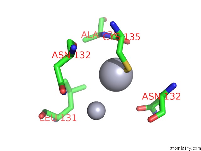
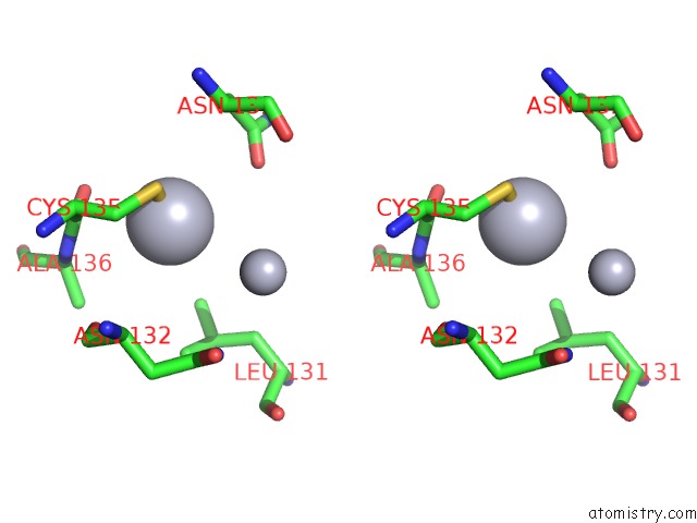
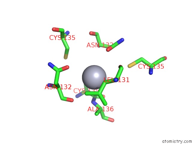
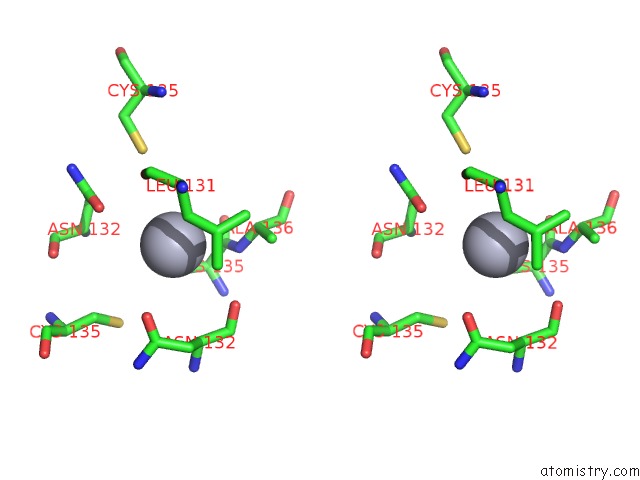
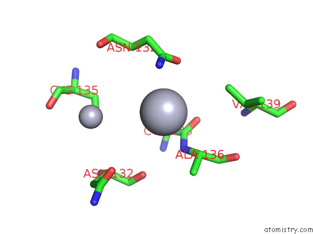
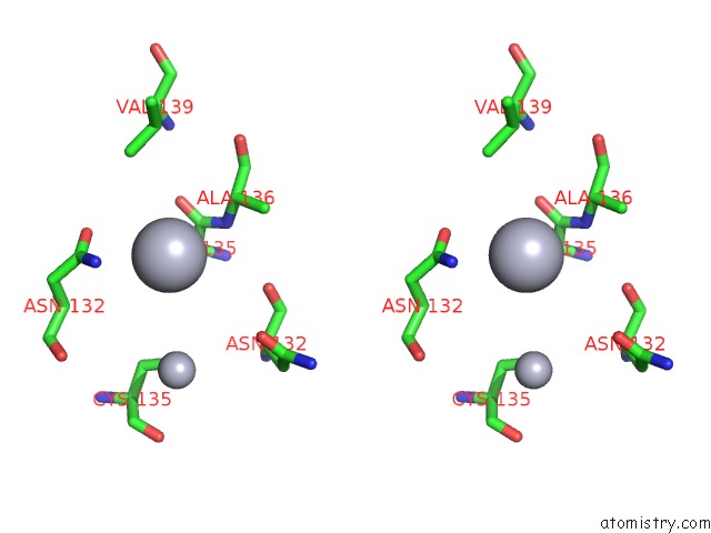
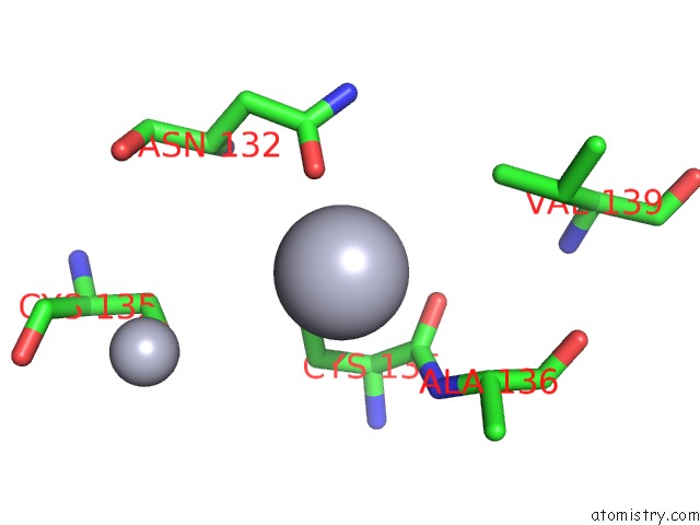
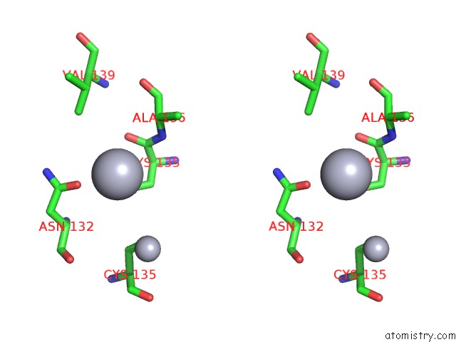
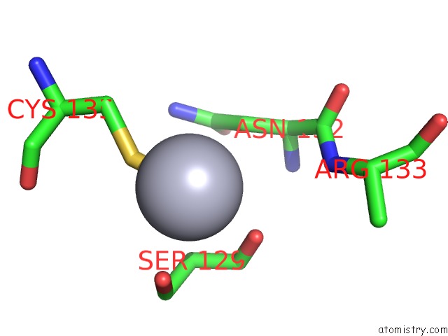
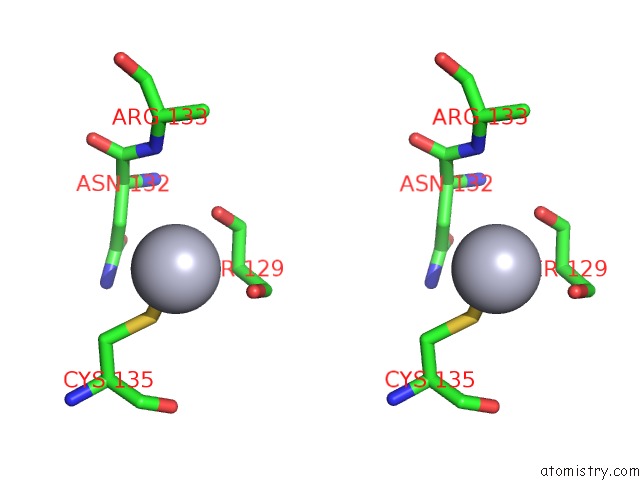
Mercury binding site 1 out of 11 in 6gbp
Go back to
Mercury binding site 1 out
of 11 in the Crystal Structure of the Oligomerization Domain of VP35 From Ebola Virus, Mercury Derivative
Mono view
Stereo pair view
Mono view
Stereo pair view
A full contact list of Mercury with other atoms in the Hg binding
site number 1 of Crystal Structure of the Oligomerization Domain of VP35 From Ebola Virus, Mercury Derivative within 5.0Å range:
|
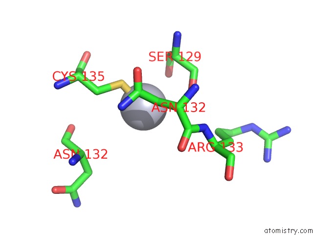
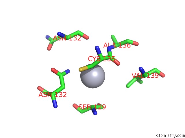
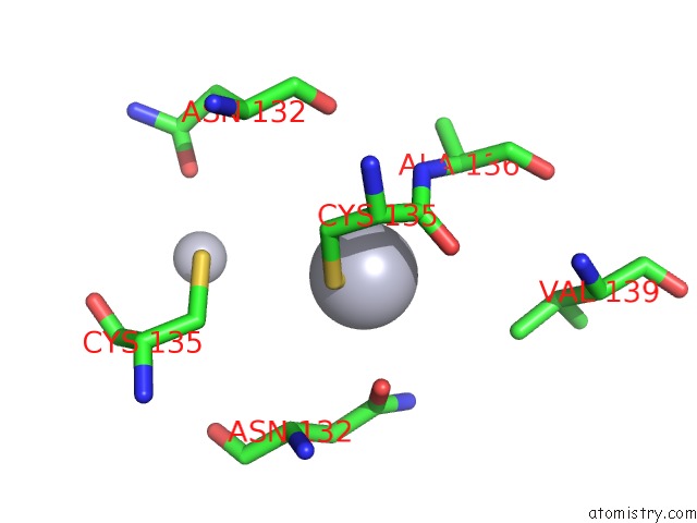
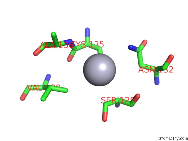
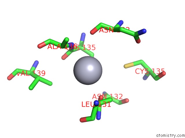
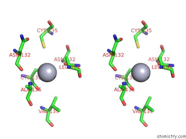
Mercury binding site 2 out of 11 in 6gbp
Go back to
Mercury binding site 2 out
of 11 in the Crystal Structure of the Oligomerization Domain of VP35 From Ebola Virus, Mercury Derivative
Mono view
Stereo pair view
Mono view
Stereo pair view
A full contact list of Mercury with other atoms in the Hg binding
site number 2 of Crystal Structure of the Oligomerization Domain of VP35 From Ebola Virus, Mercury Derivative within 5.0Å range:
|
Mercury binding site 3 out of 11 in 6gbp
Go back to
Mercury binding site 3 out
of 11 in the Crystal Structure of the Oligomerization Domain of VP35 From Ebola Virus, Mercury Derivative
Mono view
Stereo pair view
Mono view
Stereo pair view
A full contact list of Mercury with other atoms in the Hg binding
site number 3 of Crystal Structure of the Oligomerization Domain of VP35 From Ebola Virus, Mercury Derivative within 5.0Å range:
|
Mercury binding site 4 out of 11 in 6gbp
Go back to
Mercury binding site 4 out
of 11 in the Crystal Structure of the Oligomerization Domain of VP35 From Ebola Virus, Mercury Derivative
Mono view
Stereo pair view
Mono view
Stereo pair view
A full contact list of Mercury with other atoms in the Hg binding
site number 4 of Crystal Structure of the Oligomerization Domain of VP35 From Ebola Virus, Mercury Derivative within 5.0Å range:
|
Mercury binding site 5 out of 11 in 6gbp
Go back to
Mercury binding site 5 out
of 11 in the Crystal Structure of the Oligomerization Domain of VP35 From Ebola Virus, Mercury Derivative
Mono view
Stereo pair view
Mono view
Stereo pair view
A full contact list of Mercury with other atoms in the Hg binding
site number 5 of Crystal Structure of the Oligomerization Domain of VP35 From Ebola Virus, Mercury Derivative within 5.0Å range:
|
Mercury binding site 6 out of 11 in 6gbp
Go back to
Mercury binding site 6 out
of 11 in the Crystal Structure of the Oligomerization Domain of VP35 From Ebola Virus, Mercury Derivative
Mono view
Stereo pair view
Mono view
Stereo pair view
A full contact list of Mercury with other atoms in the Hg binding
site number 6 of Crystal Structure of the Oligomerization Domain of VP35 From Ebola Virus, Mercury Derivative within 5.0Å range:
|
Mercury binding site 7 out of 11 in 6gbp
Go back to
Mercury binding site 7 out
of 11 in the Crystal Structure of the Oligomerization Domain of VP35 From Ebola Virus, Mercury Derivative
Mono view
Stereo pair view
Mono view
Stereo pair view
A full contact list of Mercury with other atoms in the Hg binding
site number 7 of Crystal Structure of the Oligomerization Domain of VP35 From Ebola Virus, Mercury Derivative within 5.0Å range:
|
Mercury binding site 8 out of 11 in 6gbp
Go back to
Mercury binding site 8 out
of 11 in the Crystal Structure of the Oligomerization Domain of VP35 From Ebola Virus, Mercury Derivative
Mono view
Stereo pair view
Mono view
Stereo pair view
A full contact list of Mercury with other atoms in the Hg binding
site number 8 of Crystal Structure of the Oligomerization Domain of VP35 From Ebola Virus, Mercury Derivative within 5.0Å range:
|
Mercury binding site 9 out of 11 in 6gbp
Go back to
Mercury binding site 9 out
of 11 in the Crystal Structure of the Oligomerization Domain of VP35 From Ebola Virus, Mercury Derivative
Mono view
Stereo pair view
Mono view
Stereo pair view
A full contact list of Mercury with other atoms in the Hg binding
site number 9 of Crystal Structure of the Oligomerization Domain of VP35 From Ebola Virus, Mercury Derivative within 5.0Å range:
|
Mercury binding site 10 out of 11 in 6gbp
Go back to
Mercury binding site 10 out
of 11 in the Crystal Structure of the Oligomerization Domain of VP35 From Ebola Virus, Mercury Derivative
Mono view
Stereo pair view
Mono view
Stereo pair view
A full contact list of Mercury with other atoms in the Hg binding
site number 10 of Crystal Structure of the Oligomerization Domain of VP35 From Ebola Virus, Mercury Derivative within 5.0Å range:
|
Reference:
L.Zinzula,
I.Nagy,
M.Orsini,
E.Weyher-Stingl,
A.Bracher,
W.Baumeister.
Structures of Ebola and Reston Virus VP35 Oligomerization Domains and Comparative Biophysical Characterization in All Ebolavirus Species. Structure V. 27 39 2019.
ISSN: ISSN 1878-4186
PubMed: 30482729
DOI: 10.1016/J.STR.2018.09.009
Page generated: Sun Aug 11 07:18:10 2024
ISSN: ISSN 1878-4186
PubMed: 30482729
DOI: 10.1016/J.STR.2018.09.009
Last articles
Zn in 9MJ5Zn in 9HNW
Zn in 9G0L
Zn in 9FNE
Zn in 9DZN
Zn in 9E0I
Zn in 9D32
Zn in 9DAK
Zn in 8ZXC
Zn in 8ZUF