Mercury »
PDB 8phl-9d6a »
8phm »
Mercury in PDB 8phm: Oxalate-Bound Cobalt(II) Human Carbonic Anhydrase II
Enzymatic activity of Oxalate-Bound Cobalt(II) Human Carbonic Anhydrase II
All present enzymatic activity of Oxalate-Bound Cobalt(II) Human Carbonic Anhydrase II:
4.2.1.1; 4.2.1.69;
4.2.1.1; 4.2.1.69;
Protein crystallography data
The structure of Oxalate-Bound Cobalt(II) Human Carbonic Anhydrase II, PDB code: 8phm
was solved by
L.Gigli,
J.Malanho Silva,
L.Cerofolini,
A.L.Macedo,
C.F.G.C.Geraldes,
E.A.Suturina,
V.Calderone,
M.Fragai,
G.Parigi,
E.Ravera,
C.Luchinat,
with X-Ray Crystallography technique. A brief refinement statistics is given in the table below:
| Resolution Low / High (Å) | 26.56 / 1.45 |
| Space group | P 1 21 1 |
| Cell size a, b, c (Å), α, β, γ (°) | 41.91, 41.21, 71.71, 90, 104.29, 90 |
| R / Rfree (%) | 16.4 / 19.8 |
Other elements in 8phm:
The structure of Oxalate-Bound Cobalt(II) Human Carbonic Anhydrase II also contains other interesting chemical elements:
| Cobalt | (Co) | 1 atom |
Mercury Binding Sites:
The binding sites of Mercury atom in the Oxalate-Bound Cobalt(II) Human Carbonic Anhydrase II
(pdb code 8phm). This binding sites where shown within
5.0 Angstroms radius around Mercury atom.
In total only one binding site of Mercury was determined in the Oxalate-Bound Cobalt(II) Human Carbonic Anhydrase II, PDB code: 8phm:
In total only one binding site of Mercury was determined in the Oxalate-Bound Cobalt(II) Human Carbonic Anhydrase II, PDB code: 8phm:
Mercury binding site 1 out of 1 in 8phm
Go back to
Mercury binding site 1 out
of 1 in the Oxalate-Bound Cobalt(II) Human Carbonic Anhydrase II
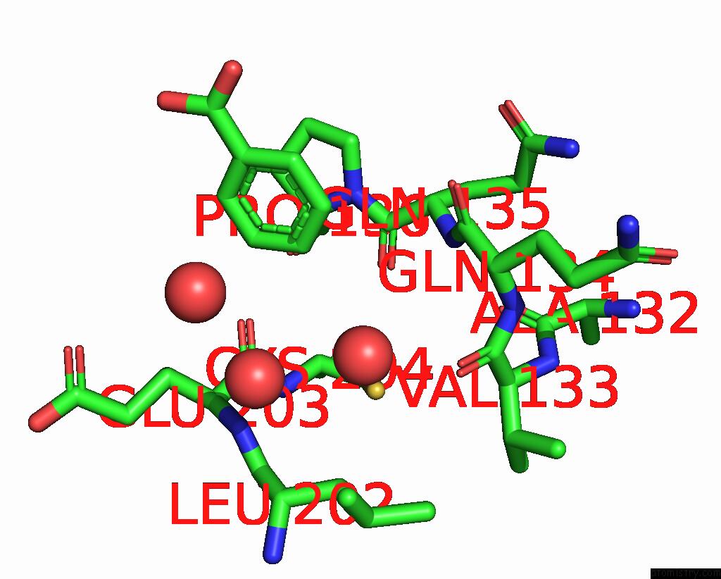
Mono view
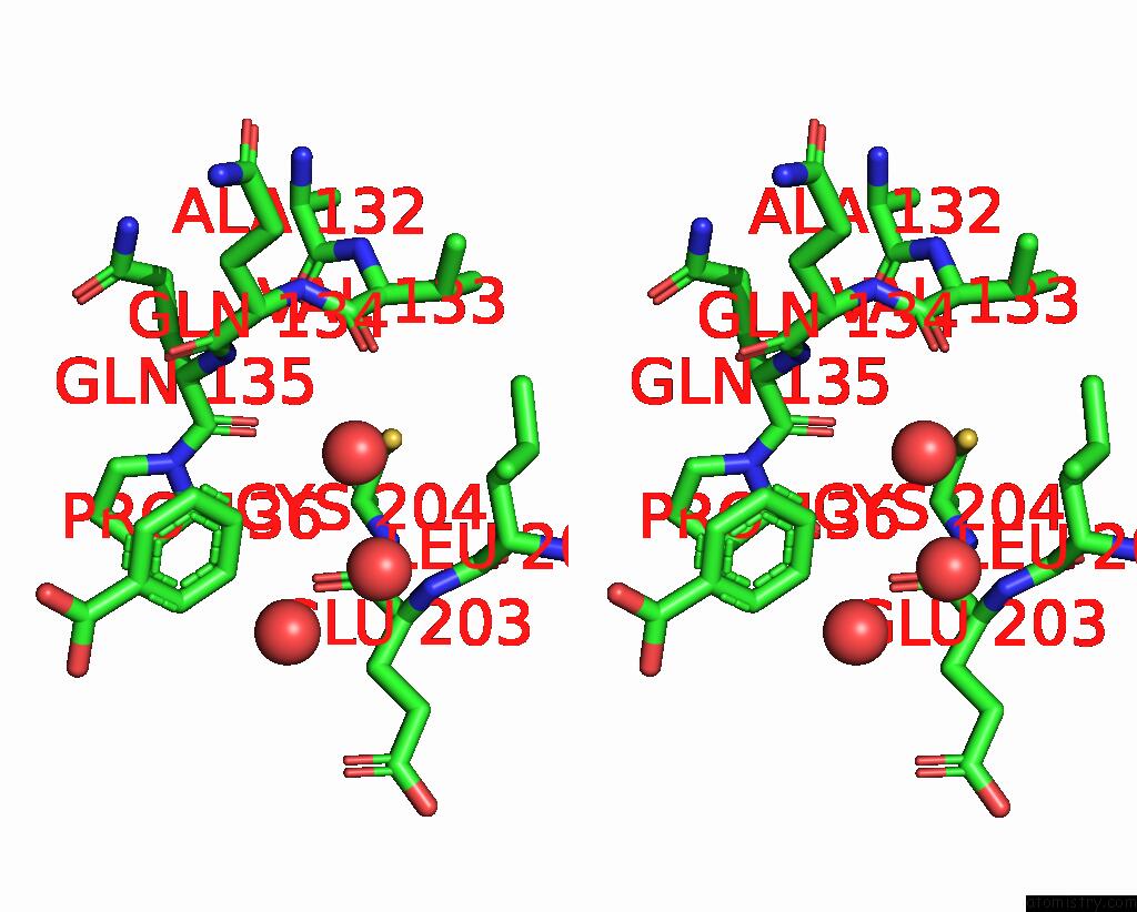
Stereo pair view
Mono view
Stereo pair view
A full contact list of Mercury with other atoms in the Hg binding
site number 1 of Oxalate-Bound Cobalt(II) Human Carbonic Anhydrase II within 5.0Å range:
|
Reference:
L.Gigli,
J.M.Silva,
L.Cerofolini,
A.L.Macedo,
C.F.G.C.Geraldes,
E.A.Suturina,
V.Calderone,
M.Fragai,
G.Parigi,
E.Ravera,
C.Luchinat.
Machine Learning-Enhanced Quantum Chemistry-Assisted Refinement of the Active Site Structure of Metalloproteins. Inorg.Chem. V. 63 10713 2024.
ISSN: ISSN 0020-1669
PubMed: 38805564
DOI: 10.1021/ACS.INORGCHEM.4C01274
Page generated: Thu Oct 31 21:27:09 2024
ISSN: ISSN 0020-1669
PubMed: 38805564
DOI: 10.1021/ACS.INORGCHEM.4C01274
Last articles
Zn in 9MJ5Zn in 9HNW
Zn in 9G0L
Zn in 9FNE
Zn in 9DZN
Zn in 9E0I
Zn in 9D32
Zn in 9DAK
Zn in 8ZXC
Zn in 8ZUF