Mercury »
PDB 12ca-1czm »
1cc8 »
Mercury in PDB 1cc8: Crystal Structure of the ATX1 Metallochaperone Protein
Protein crystallography data
The structure of Crystal Structure of the ATX1 Metallochaperone Protein, PDB code: 1cc8
was solved by
A.C.Rosenzweig,
D.L.Huffman,
M.Y.R.A.Pufahl,
T.V.O.Hou,
A.K.Wernimont,
with X-Ray Crystallography technique. A brief refinement statistics is given in the table below:
| Resolution Low / High (Å) | 50.00 / 1.02 |
| Space group | C 1 2 1 |
| Cell size a, b, c (Å), α, β, γ (°) | 56.650, 29.600, 40.770, 90.00, 114.83, 90.00 |
| R / Rfree (%) | 14.6 / 17.2 |
Mercury Binding Sites:
The binding sites of Mercury atom in the Crystal Structure of the ATX1 Metallochaperone Protein
(pdb code 1cc8). This binding sites where shown within
5.0 Angstroms radius around Mercury atom.
In total only one binding site of Mercury was determined in the Crystal Structure of the ATX1 Metallochaperone Protein, PDB code: 1cc8:
In total only one binding site of Mercury was determined in the Crystal Structure of the ATX1 Metallochaperone Protein, PDB code: 1cc8:
Mercury binding site 1 out of 1 in 1cc8
Go back to
Mercury binding site 1 out
of 1 in the Crystal Structure of the ATX1 Metallochaperone Protein
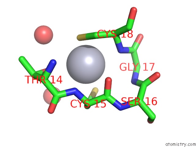
Mono view
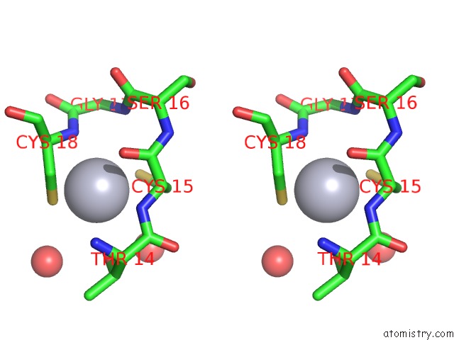
Stereo pair view
Mono view
Stereo pair view
A full contact list of Mercury with other atoms in the Hg binding
site number 1 of Crystal Structure of the ATX1 Metallochaperone Protein within 5.0Å range:
|
Reference:
A.C.Rosenzweig,
D.L.Huffman,
M.Y.Hou,
A.K.Wernimont,
R.A.Pufahl,
T.V.O'halloran.
Crystal Structure of the ATX1 Metallochaperone Protein at 1.02 A Resolution. Structure Fold.Des. V. 7 605 1999.
ISSN: ISSN 0969-2126
PubMed: 10404590
DOI: 10.1016/S0969-2126(99)80082-3
Page generated: Sat Aug 10 23:22:48 2024
ISSN: ISSN 0969-2126
PubMed: 10404590
DOI: 10.1016/S0969-2126(99)80082-3
Last articles
Zn in 9MJ5Zn in 9HNW
Zn in 9G0L
Zn in 9FNE
Zn in 9DZN
Zn in 9E0I
Zn in 9D32
Zn in 9DAK
Zn in 8ZXC
Zn in 8ZUF